Patrick Korf1, Wolfgang Thaiss2, Ambros J. Beer2, Meinrad Beer3, Dominik Nickel1, and Thomas Vahle1
1Siemens Healthcare GmbH, Erlangen, Germany, 2Department of Nuclear Medicine, University Hospital Ulm, Ulm, Germany, 3Department of Diagnostic and Interventional Radiology, University Hospital Ulm, Ulm, Germany
1Siemens Healthcare GmbH, Erlangen, Germany, 2Department of Nuclear Medicine, University Hospital Ulm, Ulm, Germany, 3Department of Diagnostic and Interventional Radiology, University Hospital Ulm, Ulm, Germany
We developed
a free-breathing attenuation correction method based on 2pt Dixon for
whole-body PET/MR exams. In addition to the attenuation map, high resolution
Dixon images are generated. Initial results are presented.
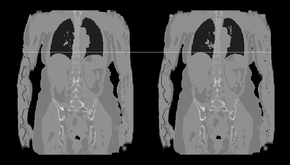
Figure 1: Comparison
of whole-body attenuation maps of a healthy volunteer. Left: Breath-hold
protocol. Right: Free-breathing protocol. The first two bed positions were
acquired in breath-hold and free-breathing respectively. The last bed position
was acquired using the breath-hold protocol without breath-hold command. In
both cases the attenuation map consisting of five compartments (air, lung
tissue, soft tissue, fat tissue and bone) could be generated successfully. The truncated
arms were recovered as well as described in [2].

Figure 2: Comparison of Dixon image quality. Left Column:
Dixon water, acquisition with breath-hold protocol. Right Column: Dixon water,
free-breathing acquisition. Free-breathing images show a more distinct boundary
of the liver dome and appear to be sharper in the coronal and sagittal
reformats (middle and bottom rows). The image quality in the axial plane (top
row) is comparable. Images were acquired after a clinical study, Gadovist was
given as part of the scan protocol prior to the shown acquisitions.