Bo Peng1,2,3, Baohua Hu1,2,3, Mao Sheng4, Yuqi Liu4, Zhongchang Miao5, Zijun Dong6, Jian Bao7, SiSeung Kim7, Bing Keong Li7, and Yakang Dai1,2,3
1Suzhou Institute of Biomedical Engineering and Technology, Chinese Academy of Sciences, Suzhou, China, 2Suzhou Key Laboratory of Medical and Health Information Technology, Suzhou, China, 3Jinan Guoke Medical Engineering Technology Development co., Ltd., Jinan, China, 4Department of Radiology, Children’s Hospital of Soochow University, Suzhou, China, 5Department of Radiology, The First People’s Hospital of Lianyungang, Jiangsu Province, China, 6Department of Medical Imaging, Lianyungang Women and Children Hospital and Health Institute, Jiangsu Province, China, 7Jiangsu LiCi Medical Device Co., Ltd., Lianyungang, China
1Suzhou Institute of Biomedical Engineering and Technology, Chinese Academy of Sciences, Suzhou, China, 2Suzhou Key Laboratory of Medical and Health Information Technology, Suzhou, China, 3Jinan Guoke Medical Engineering Technology Development co., Ltd., Jinan, China, 4Department of Radiology, Children’s Hospital of Soochow University, Suzhou, China, 5Department of Radiology, The First People’s Hospital of Lianyungang, Jiangsu Province, China, 6Department of Medical Imaging, Lianyungang Women and Children Hospital and Health Institute, Jiangsu Province, China, 7Jiangsu LiCi Medical Device Co., Ltd., Lianyungang, China
Low-field MRI is
foreseeable as a safer system for infants. We developed an automated image
processing method. It is also capable to
automatically construct the surfaces of the cerebral
cortex and provides automatic quantitative analysis of selected region of
interest.
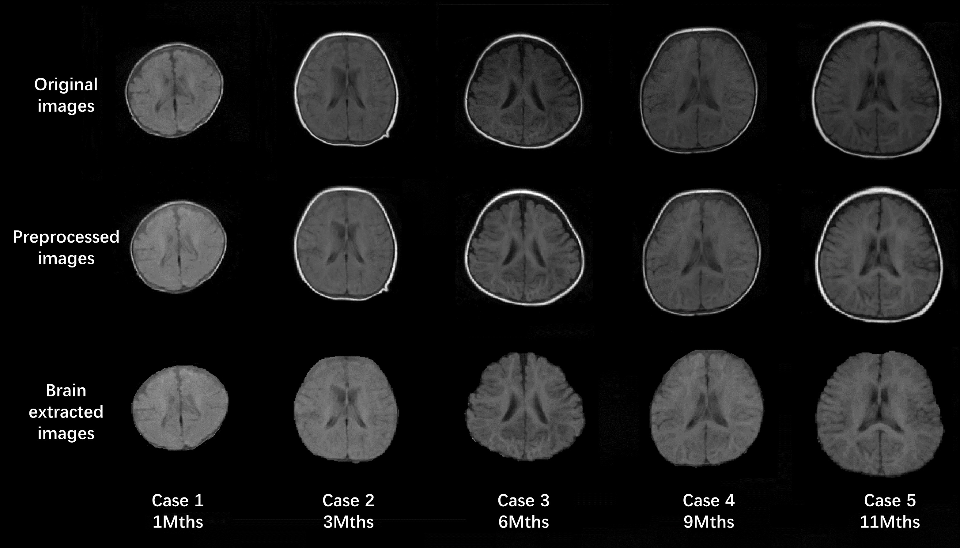
Figure 2. Original,
preprocessed, extracted brain images of infants at various age month. Top row shows the original T1W images at 0.35T. Middle row shows
preprocessed images after de-nosing and N3 bias correction. Bottom row shows
the extracted brain with the skull removed.
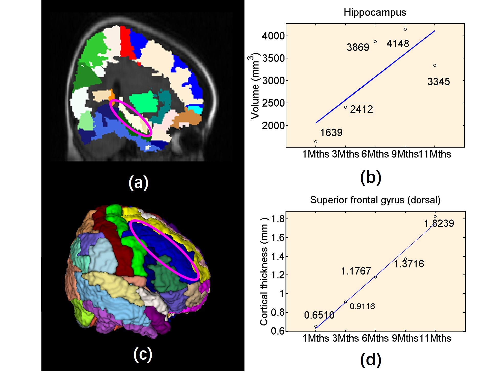
Figure
5. Quantitative calculation results of ROI volume, cortical thickness after
brain labeling. (a) Automated labeling results on voxel-wise image
(hippocampus). (b) Quantitative calculation of hippocampus volume for five
cases. (c) Labeling ROIs on cortical surface on surface-based (superior frontal
gyrus (dorsal)). (d) Cortical thickness for superior frontal gyrus (dorsal) of
different cases.