Sophie Malaquin1, Eloïse Mougel1, Melissa Vincent1, and Julien Valette1
1Université Paris-Saclay, Commissariat à l'Energie Atomique et aux Energies Alternatives (CEA), Centre National de la Recherche Scientifique (CNRS), Molecular Imaging Research Center (MIRCen), Laboratoire des Maladies Neurodégénératives, Fontenay aux Roses, France
1Université Paris-Saclay, Commissariat à l'Energie Atomique et aux Energies Alternatives (CEA), Centre National de la Recherche Scientifique (CNRS), Molecular Imaging Research Center (MIRCen), Laboratoire des Maladies Neurodégénératives, Fontenay aux Roses, France
In an effort to optimize lactate detection in diffusion-weighted
NMR spectroscopy, we compare different sequences including a spin echo sequence
using polychromatic pulse to suppress the J-coupling. This sequence results in
stronger lactate signal and more precise diffusion measurements.
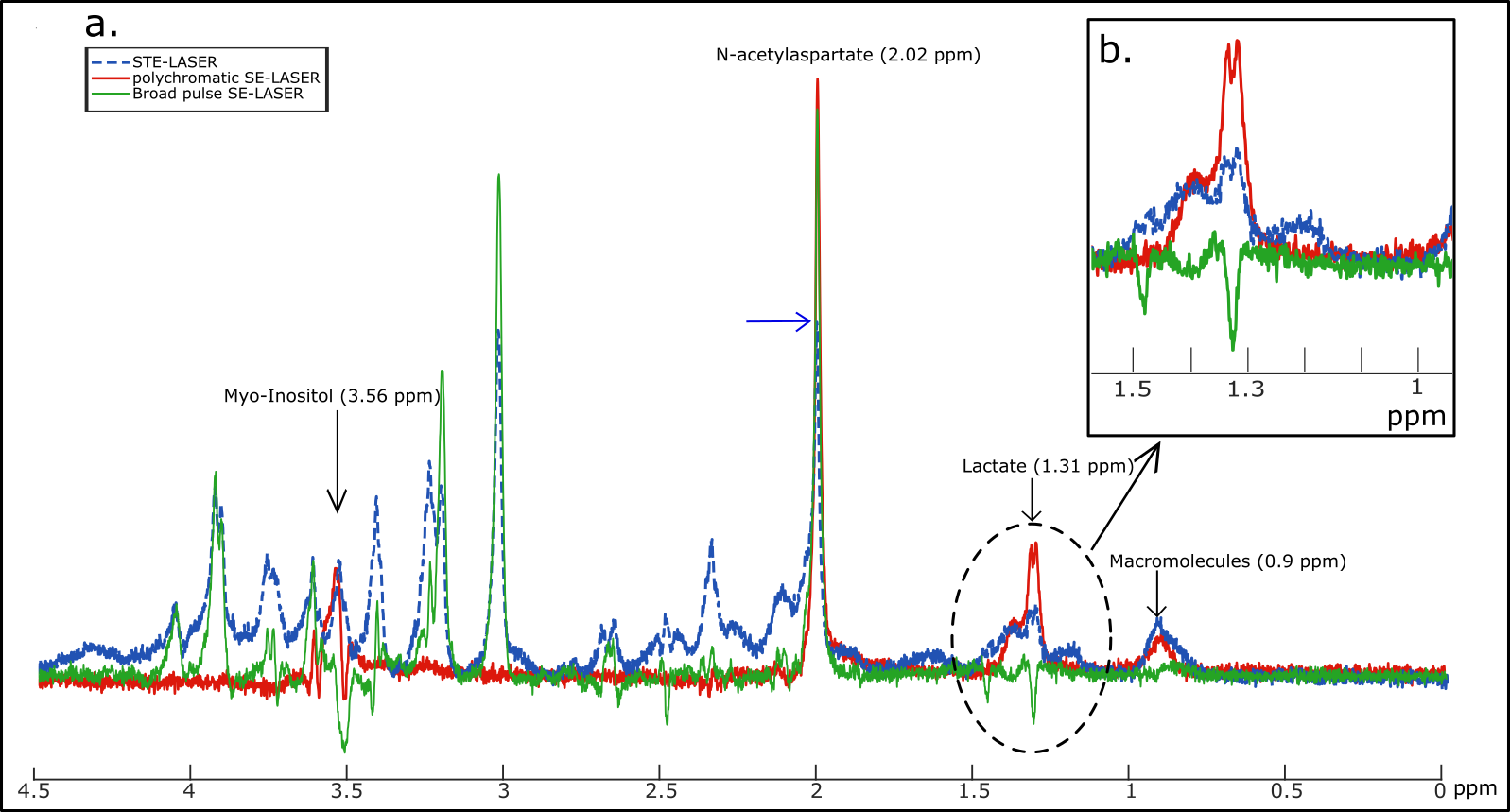
Figure 2: a. Spectra without
diffusion-weighting obtained with the STE-LASER sequence (blue dotted line),
the broad pulse SE-LASER sequence (green line) and the polychromatic SE-LASER
sequence (red line). b. The gain of lactate signal is spectacular when using
the polychromatic SE-LASER sequence.
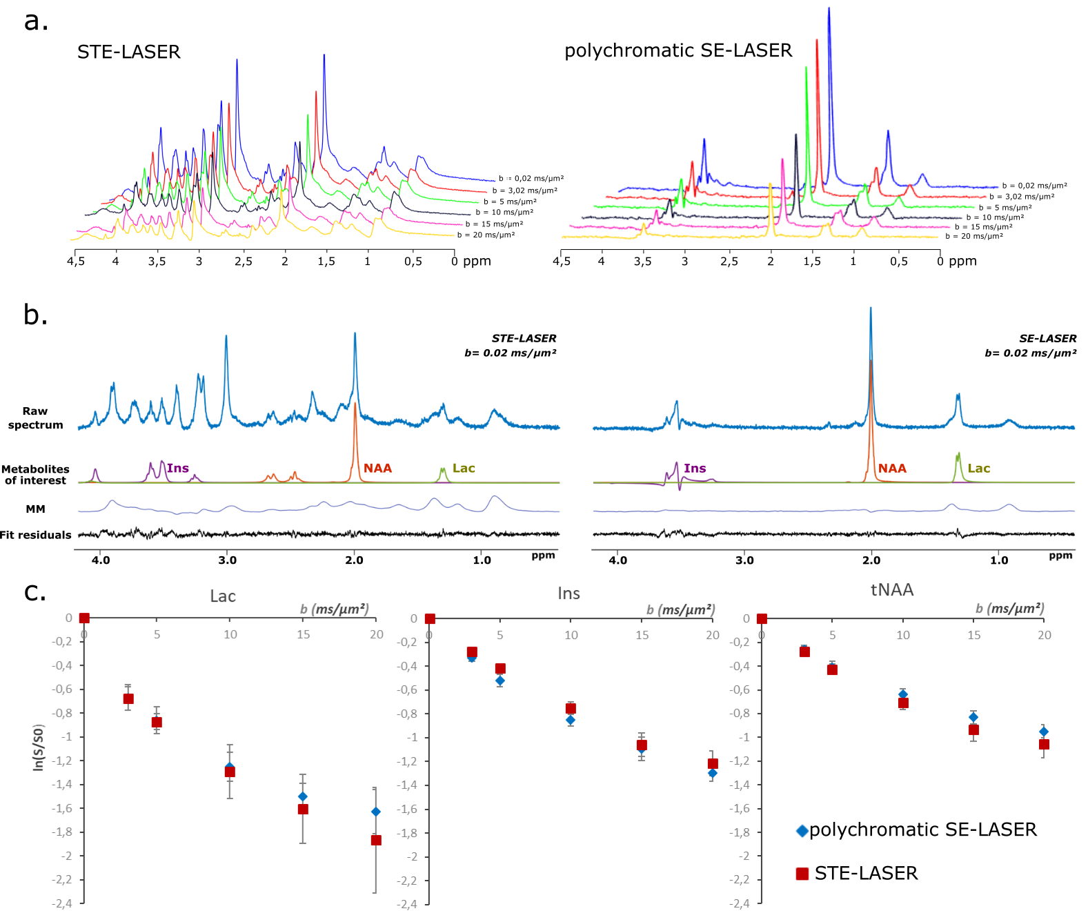
Figure 3: STE-LASER and
polychromatic SE-LASER data at high diffusion-weighting in WT mice. a. Spectra
acquired up to high diffusion-weighting (from b = 0.02 ms/µm² to 20 ms/µm²)
with the STE-LASER sequence (left) and the polychromatic SE-LASER sequence
(right).
b. LCModel analysis for
metabolites of interest. Macromolecules contribution on 1.31 ppm lactate peak
is noticeable.
c. Signal attenuation as a
function of b for three metabolites by using the two sequences (blue diamonds =
polychromatic SE-LASER, red squares = STE-LASER). Data points and error bars
stand for mean ± s.d.