Jullie W Pan1, Victor W Yushmanov2, Chan H Moon3, Brian Soher4, Frank H Lieberman5, and Hoby P Hetherington2
1Radiology, University of Pittsburgh, Pittsburgh, PA, United States, 2university of pittsburgh, pittsburgh, PA, United States, 3University of Pittsburgh, Pittsburgh, PA, United States, 4Radiiology, Duke University, Durham, NC, United States, 5Neurology, University of Pittsburgh, Pittsburgh, PA, United States
1Radiology, University of Pittsburgh, Pittsburgh, PA, United States, 2university of pittsburgh, pittsburgh, PA, United States, 3University of Pittsburgh, Pittsburgh, PA, United States, 4Radiiology, Duke University, Durham, NC, United States, 5Neurology, University of Pittsburgh, Pittsburgh, PA, United States
Spectral quality of fast 7T MR spectroscopic imaging is evaluated for multiplet compounds including Glutamine, myo-Inositol, Choline and demonstrated in brain tumor patients
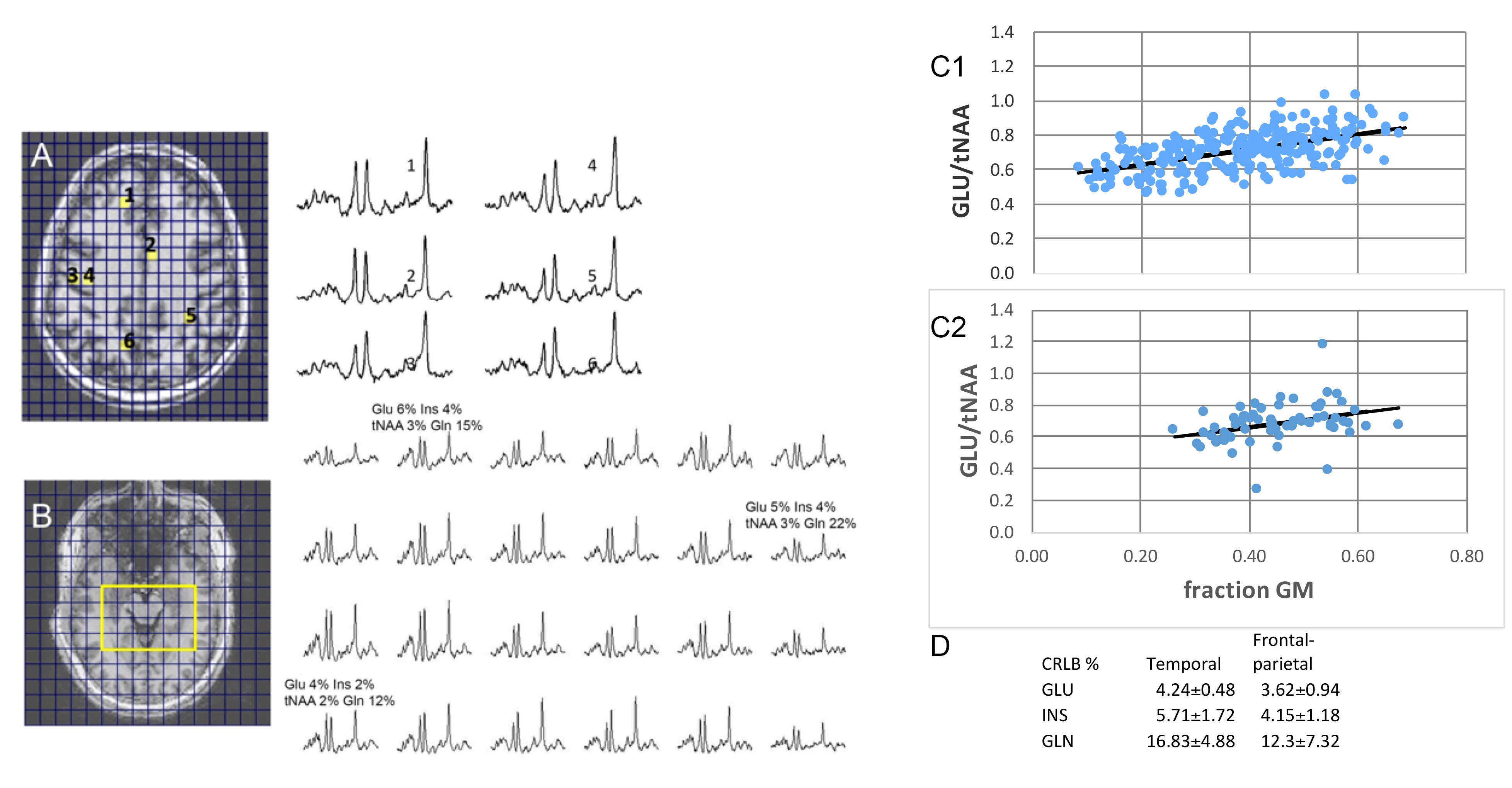
FIg. 2. (A,B) Spectral data from the frontal-parietal (A) and temporal (B) regions. CRLB % are shown for several loci in the temporal study. (C1,C2) The GLU/tNAA regresses significantly with fraction GM from both studied regions. (D) The CRLB % are shown for GLU, INS and GLN for the frontal-parietal and temporal regions.
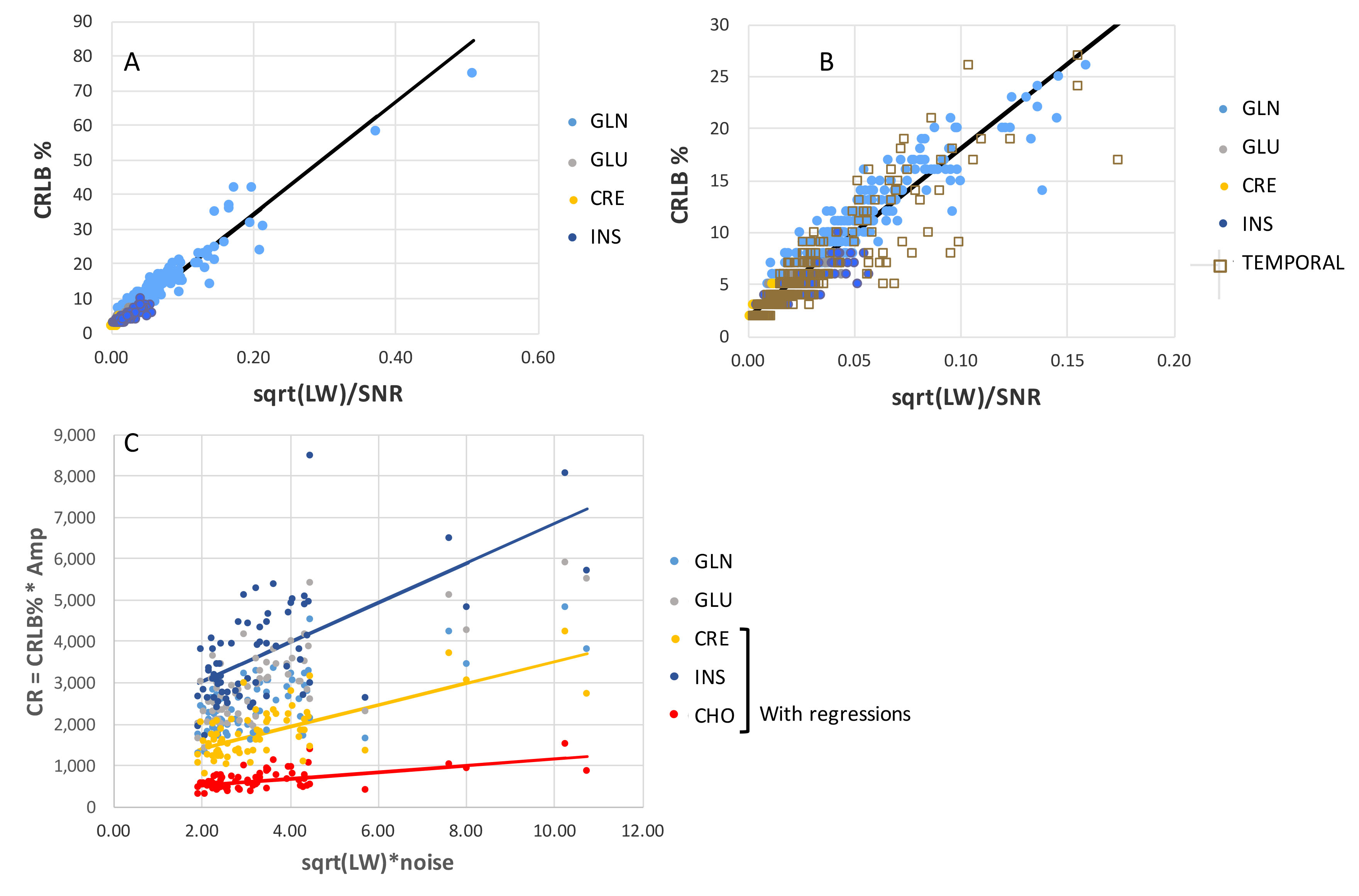
Fig. 3. (A) Regression of CRLB % with ξk=√LW/SNRk from the frontal-parietal region. (B) Same as (A) but with superimposed data from the temporal region, and magnified in X and Y axes. (C) Plots of CRk = CRLBk% * Ampk, with ξ0 =√LW*σ, with different compounds. The regressions for CRE, INS and CHO are shown.