Ralf Mekle1, Jochen B. Fiebach1, and Heiner Stuke2
1Center for Stroke Research Berlin, Charité – Universitätsmedizin Berlin, Berlin, Germany, 2Department of Psychiatry and Psychotherapy, Charité – Universitätsmedizin Berlin, Berlin, Germany
1Center for Stroke Research Berlin, Charité – Universitätsmedizin Berlin, Berlin, Germany, 2Department of Psychiatry and Psychotherapy, Charité – Universitätsmedizin Berlin, Berlin, Germany
Combining fMRI and 1H MRS
at 3T revealed that reduced activation in the fusiform face area is related to higher hallucination
proneness and lower glutamate levels, thus supporting theories of impaired
glutamatergic transmission being involved in the formation of hallucinations.
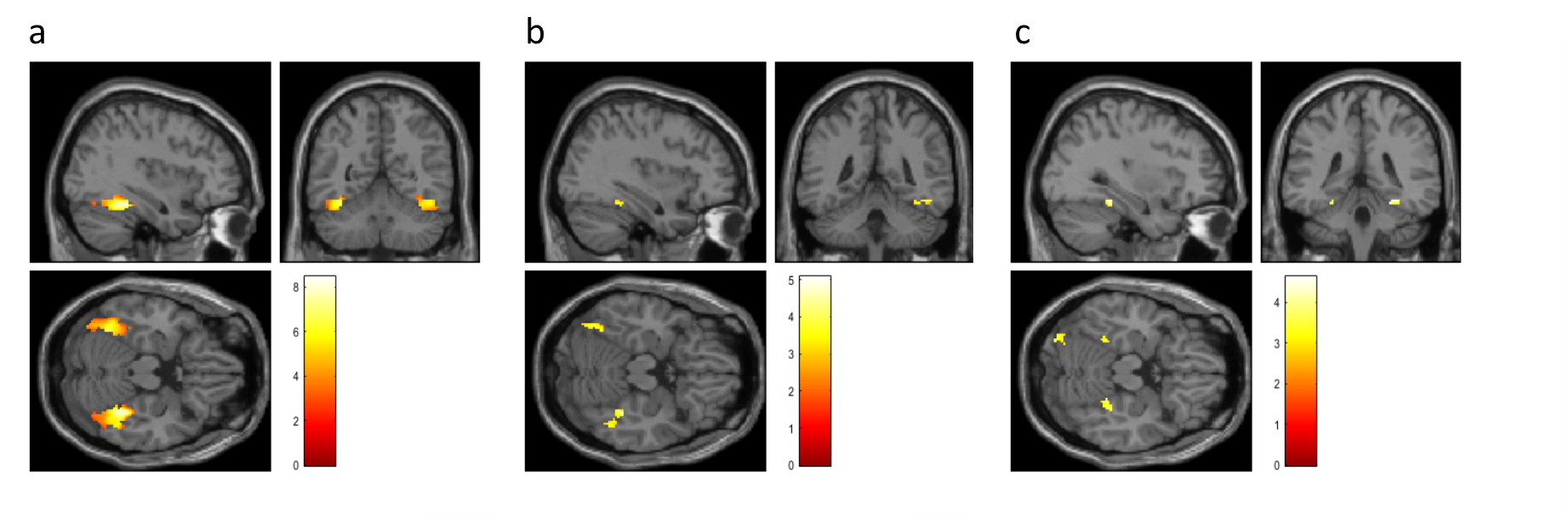
Fig. 2. Activations in the
a priori defined ROI of the bilateral gyrus fusisormis for face-stimuli versus
non-face stimuli (a, N = 18, MNI peak voxel = 38, -38, -18, puncorr < 0.001, pfwe < 0.001) and correlations of
these activations with hallucination proneness in the face task (b, negative
correlation, N = 18, MNI peak voxel = 38, -38, -18, puncorr < 0.001, pfwe = 0.040) and glutamate/NAA
ratios obtained from MRS (c, N = 16, MNI peak voxel = 32, -40, -16, puncorr < 0.001, pfwe = 0.121). In the figure, p-values of 0.005 were used for
illustration purposes.
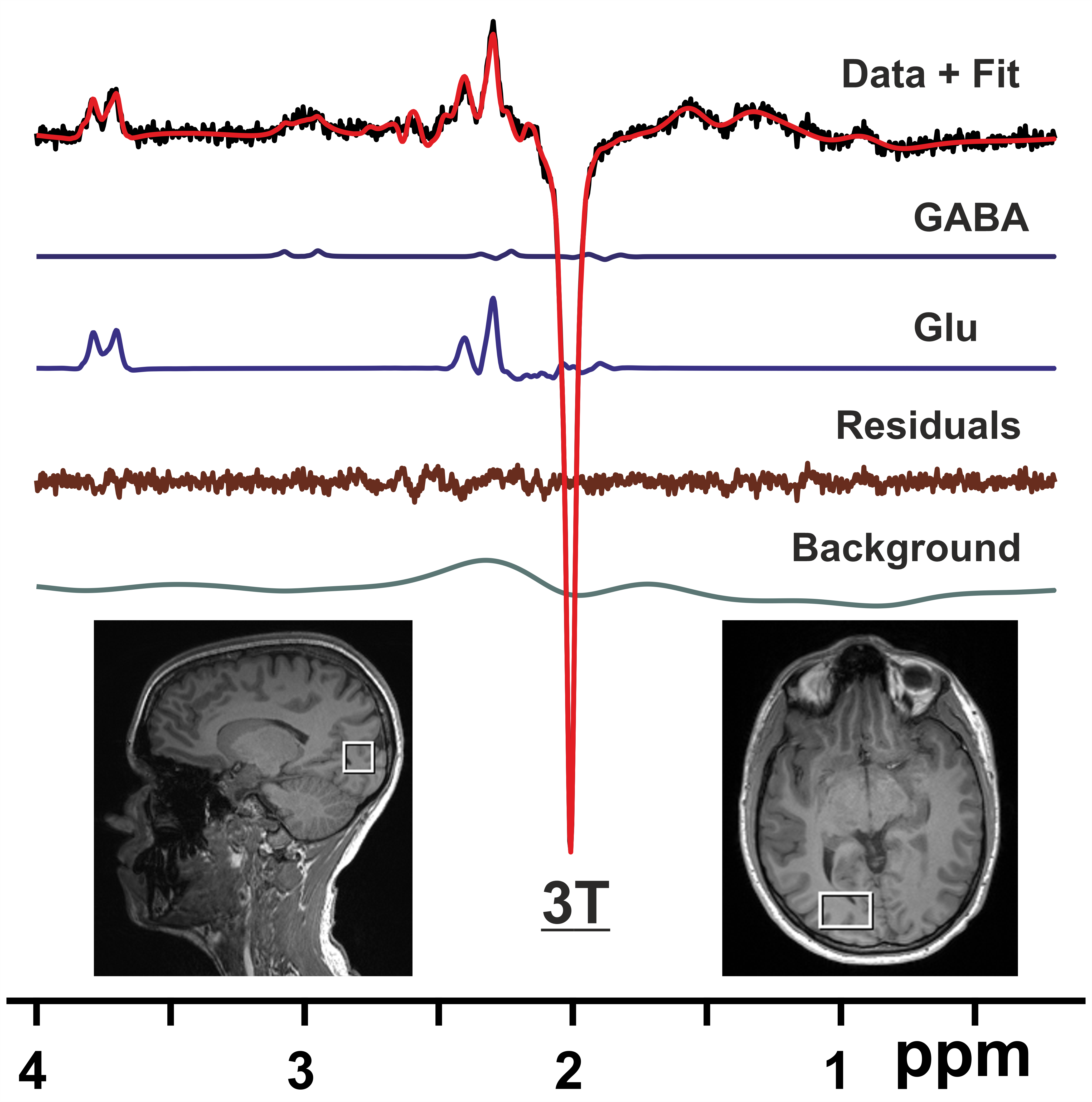
Fig. 1. 1H difference MR spectra from a VOI
in the right visual cortex (bottom inset) acquired at 3T with the MEGA-PRESS
sequence (TR/TE = 3000/68 ms) for a healthy volunteer together with
LCModel fit, LCModel output for GABA, Glu, fit residuals, and background.
Preprocessing of data included coil combination, removal of bad averages, and
phase and frequency correction.