Graeme A. Keith1, Amir Seginer2, David A. Porter1, and Rita Schmidt3
1Institute of Neuroscience and Psychology, University of Glasgow, Glasgow, Scotland, 2Siemens Healthcare Ltd, Rosh Ha’ayin, Israel, 3Neurobiology Department, Weizmann Institute of Science, Rehovot, Israel
1Institute of Neuroscience and Psychology, University of Glasgow, Glasgow, Scotland, 2Siemens Healthcare Ltd, Rosh Ha’ayin, Israel, 3Neurobiology Department, Weizmann Institute of Science, Rehovot, Israel
Readout-segmented COKE achieves high spectral width EPSI spectra,
with coherent phase evolution at 7T, in the human brain. Residual lipid
contamination, due to the readout-segmented trajectory, is investigated in
simulations and phantom scans.
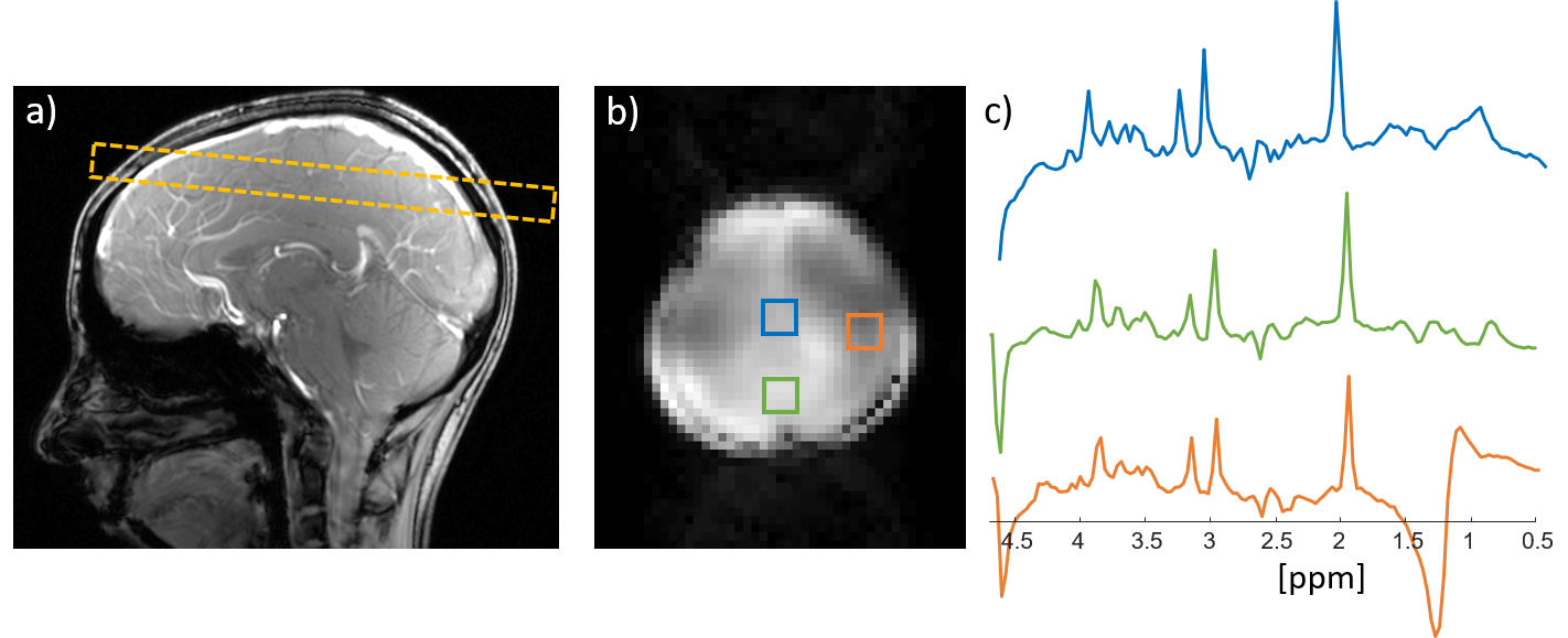
Figure
3: Human
scan results. a) Reference image with the scan location, b) water image with
the same parameters as the spectroscopic imaging scan. c) 1H spectra
at three regions shown in b). The scan parameters are described in the main
text.
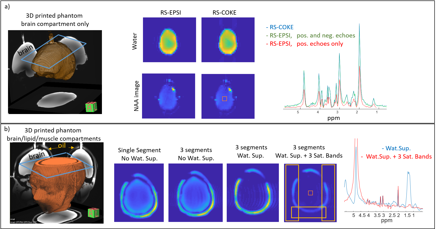
Figure
4: 3D-shaped
phantom imaging a) RS-EPSI versus RS-COKE with 3 readout segments with the “brain only” phantom. Images show water and
NAA maps and the spectrum for the central box (5x5 pixels). b) 3D-shaped
phantom including brain-like compounds and a
superficial compartment with a lipid-like compound. The slice location is
shown by blue overlay. Images compare single and 3-segment water images, a
3-segment acquisition with water suppression, and a 3-segment acquisition with
both water suppression and three saturation bands. Sequence parameters are given in the main text.