Lukas Hingerl1, Wolfgang Bogner1, Bernhard Strasser1, Petr Bednarik1, Stanislav Motyka1, Eva Heckova1, Ivica Just1, Alexandra Lipka1, Ovidiu Andronesi2, Stephan Gruber1, Siegfried Trattnig1,3, and Gilbert Hangel1,4
1Department of Biomedical Imaging and Image-guided Therapy, Medical University of Vienna, High Field MR Centre, Vienna, Austria, 2Department of Radiology, Massachusetts General Hospital, Martinos Center for Biomedical Imaging, Boston, MA, United States, 3Christian Doppler Laboratory for Clinical Molecular MR Imaging, Vienna, Austria, 4Department of Neurosurgery, Medical University of Vienna, Vienna, Austria
1Department of Biomedical Imaging and Image-guided Therapy, Medical University of Vienna, High Field MR Centre, Vienna, Austria, 2Department of Radiology, Massachusetts General Hospital, Martinos Center for Biomedical Imaging, Boston, MA, United States, 3Christian Doppler Laboratory for Clinical Molecular MR Imaging, Vienna, Austria, 4Department of Neurosurgery, Medical University of Vienna, Vienna, Austria
3D-FIDESI-MRSI
enables whole-brain lactate mapping without lipid contamination by
extending an FID sequence with a spin-echo readout, which features a
reduced but oversampled spherical k-space readout.
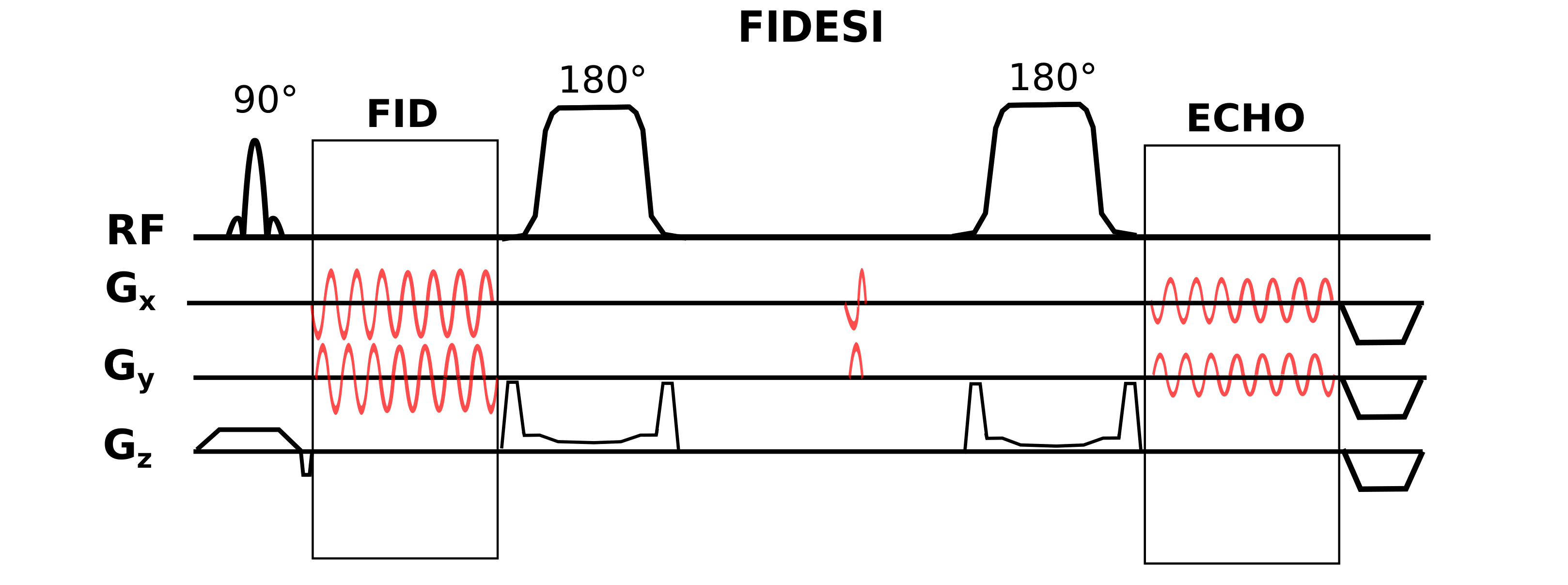
Figure 1: FIDESI features two analog-to-digital-converter readouts: Large k-space coverages for the high SNR FID scan and a small k-space coverage for the low SNR echo scan at TE 288 ms. The crusher gradient superimposed on the localization gradient ramp-down caused spectral acoustic sideband artifacts. A special pair of gradients was played out between the GOIA pulses for nulling the zero-order magnetization caused by the initial CRT gradients which move the trajectory to the specific ring radii.
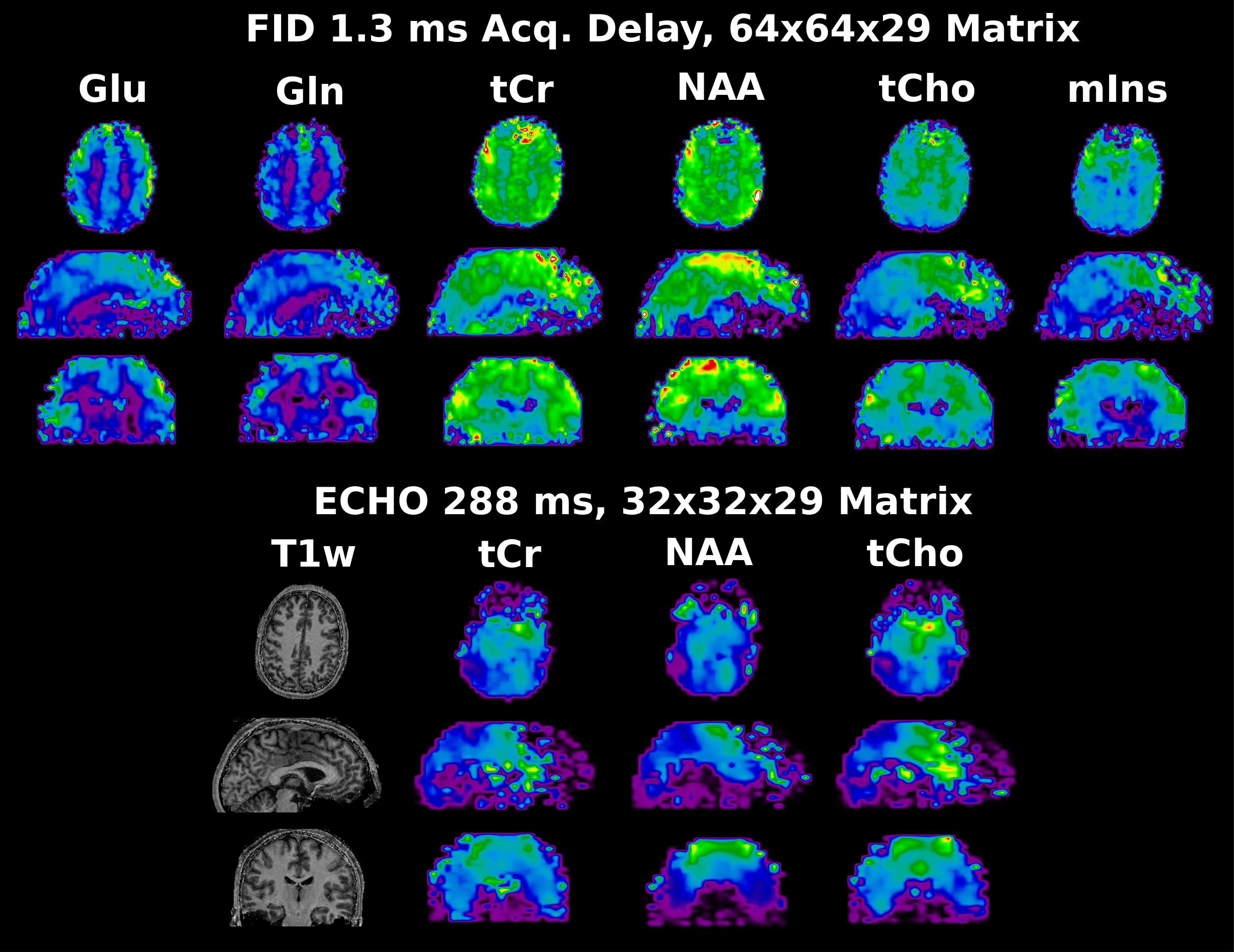
Figure 4: Whole brain metabolic Maps of the healthy Volunteer. FID maps have an effective measured matrix size of 64x64x29, echo maps have an effective measured matrix size of 32x32x15, but are interpolated to 32x32x29 before quantification. All maps are additionally interpolated by a factor of 2 for visualization purposes.