Oun Al-iedani1,2, Jeannette Lechner-Scott2,3,4, Rodney Lea2, Ovidiu Andronesi5, and Saadallah Ramadan2,6
1School of Health Sciences, University of Newcastle, Newcastle, Australia, 2Hunter Medical Research Institute, Newcastle, Australia, 3Department of Neurology, John Hunter Hospital, Newcastle, Australia, 4School of Medicine and Public Health, University of Newcastle, Newcastle, Australia, 5Harvard Medical School, Massachusetts General Hospital, Boston, MA, United States, 6Faculty of Health and Medicine, University of Newcastle, Newcastle, Australia
1School of Health Sciences, University of Newcastle, Newcastle, Australia, 2Hunter Medical Research Institute, Newcastle, Australia, 3Department of Neurology, John Hunter Hospital, Newcastle, Australia, 4School of Medicine and Public Health, University of Newcastle, Newcastle, Australia, 5Harvard Medical School, Massachusetts General Hospital, Boston, MA, United States, 6Faculty of Health and Medicine, University of Newcastle, Newcastle, Australia
Novel pipeline of Multi-slice spiral-MRSI coupled
with DMRs revealed NAA mapping in WM-lesions
was significantly lower than NAWM-MS and HCs, and is sensitive
in diagnosing NAWM in RRMS.
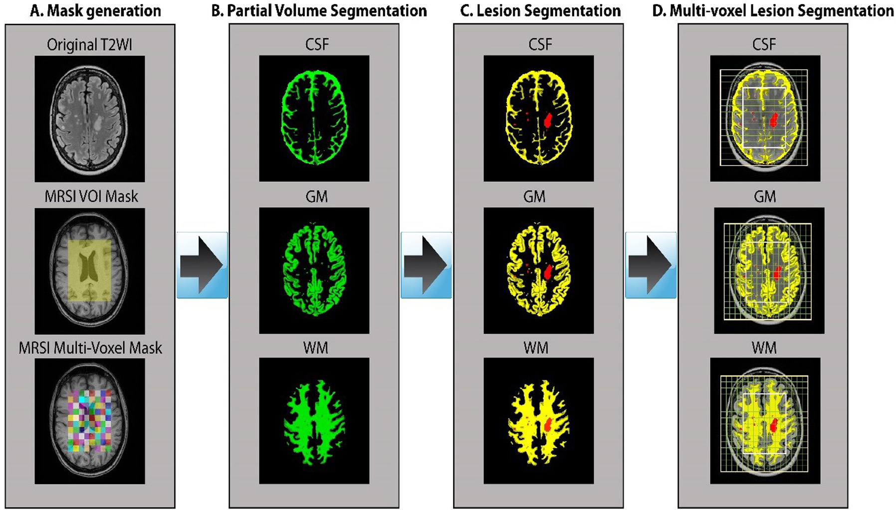
Pipeline of volumetric brain tissue segmentation from MRI and MRSI data.
A. A binary mask of a MRSI single slice VOI (8x10 cm2) was created
using the SPM toolbox. B. partial volume masks for each tissue type were
created using FSL FAST. C. Partial volume segmentation of the lesion filled
T1-MPRAGE structural images. D. Tissue segmentation of MRSI VOI (CSF, GM and
WM) and lesion segmentation overlaid on the T2-FLAIR image.
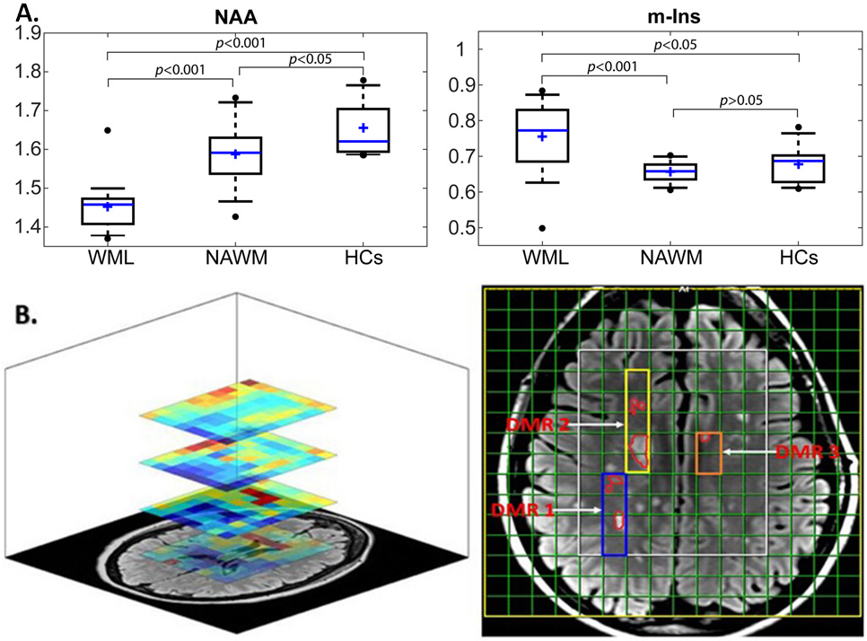
A. Box plot
of significant differences in the neurometabolic ratios (NAA/tCr)and
(m-Ins/tCr) for WML voxels, NAWM-MS and HC voxels in four slices of one DMR,
located within deep cortical white matter in posterior parietal lobes (left).
B. Mapping of NAA/tCr
ratios in single slice and multi-slice of 3 DMRs located in one axial brain
slice of an MS patient, using spiral-MRSI based representation of data, overlayed with
structural image.