Philipp Lazen1, Sahar Nassirpour2, Paul Chang2, Lukas Hingerl1, Karl Rössler3, Siegfried Trattnig1,4, Wolfgang Bogner1, and Gilbert Hangel1,3
1Department of Biomedical Imaging and Image-guided Therapy, Medical University of Vienna, Vienna, Austria, 2MR Shim GmbH, Reutlingen, Germany, 3Department of Neurosurgery, Medical University of Vienna, Vienna, Austria, 4Christian Doppler Laboratory for Clinical Molecular MR Imaging, Vienna, Austria
1Department of Biomedical Imaging and Image-guided Therapy, Medical University of Vienna, Vienna, Austria, 2MR Shim GmbH, Reutlingen, Germany, 3Department of Neurosurgery, Medical University of Vienna, Vienna, Austria, 4Christian Doppler Laboratory for Clinical Molecular MR Imaging, Vienna, Austria
A 24-channel local shim coil array was tested for 7 T 3D MRSI in healthy volunteers. The B0 field strength’s standard deviation decreased by around 20% overall and signal to noise ratios and full widths at half maximum were improved. Some preliminary MRSI data was gathered.
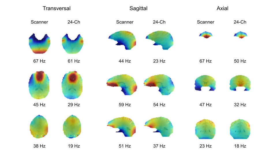
Figure 2: Representative B0 maps for both shim settings (once only with scanner shim of up to second order spherical harmonics and once with the 24-channel local shim array) are shown in three orientations overlaid on anatomical images for a single volunteer. B0 maps are plotted between [-100Hz,100Hz]. The standard deviation of the frequency shifts are reported under each corresponding B0 map.
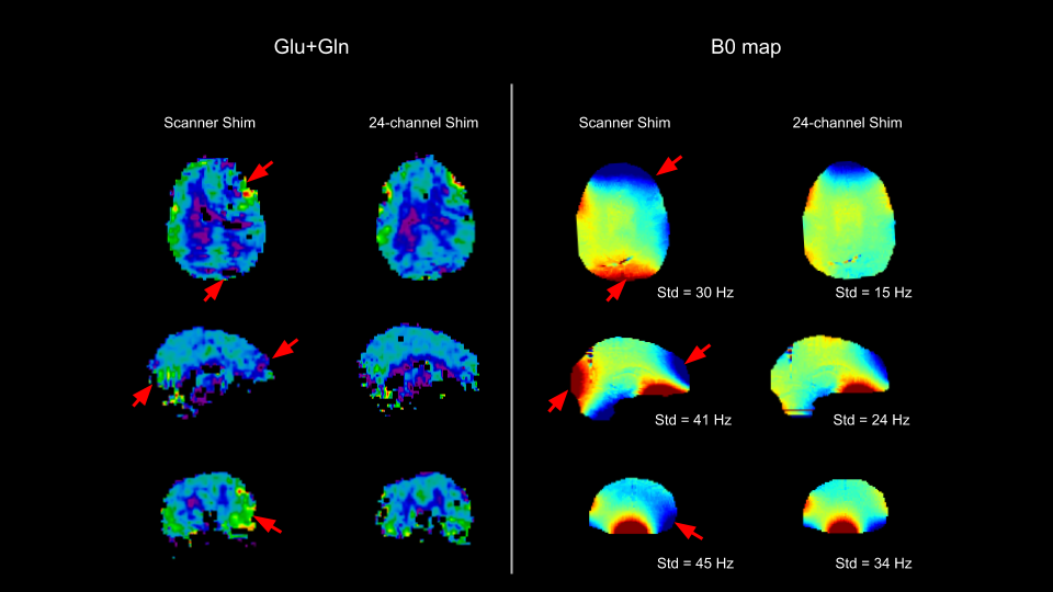
Figure 4: Representative map of glutamate and glutamine (Glu+Gln) for the standard scanner shim and the 24-channel shim, along with the corresponding B0 maps. Regions that suffer from poor B0 shimming quality with the scanner shim are marked by red arrows. The B0 maps are plotted between -60 Hz (blue) and 60 Hz (red) and the corresponding standard deviations are noted below each B0 map.