Daniel Spielman1, Phil Adamson2, Ralph Hurd1, Meng Gu1, Kirk Riemer3, Ryan Beckman3, Michael Ma3, Kenichi Okamura3, and Frank Hanley3
1Radiology, Stanford University, Stanford, CA, United States, 2Electrical Engineering, Stanford University, Stanford, CA, United States, 3Cardiothoracic Surgery, Stanford University, Stanford, CA, United States
1Radiology, Stanford University, Stanford, CA, United States, 2Electrical Engineering, Stanford University, Stanford, CA, United States, 3Cardiothoracic Surgery, Stanford University, Stanford, CA, United States
Acute brain metabolic changes were observed via 3T 1H MRS in a neonatal piglet model of cardiopulmonary bypass surgery, with the goal of finding optimal surgical parameters for minimizing brain injury. Findings include robust measures of brain temperature, hypoxia, and energy metabolism.
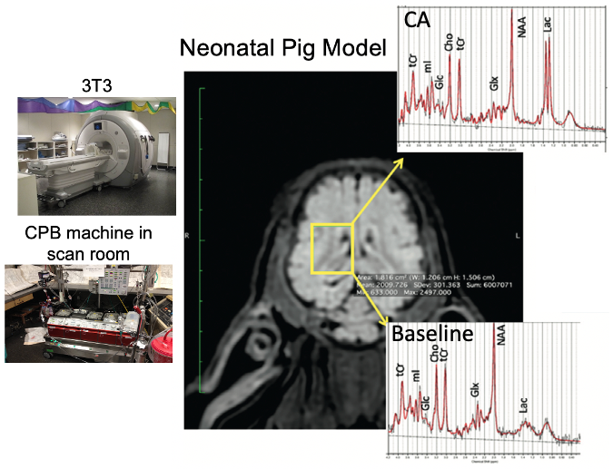
Figure 1. 3T MRI scanner setup for neonatal pig DHCA-CPB study. A coronal T1-weighted MRI with the selected MRS voxel and representative spectra at baseline and end circulatory arrest (CA) time points are shown.
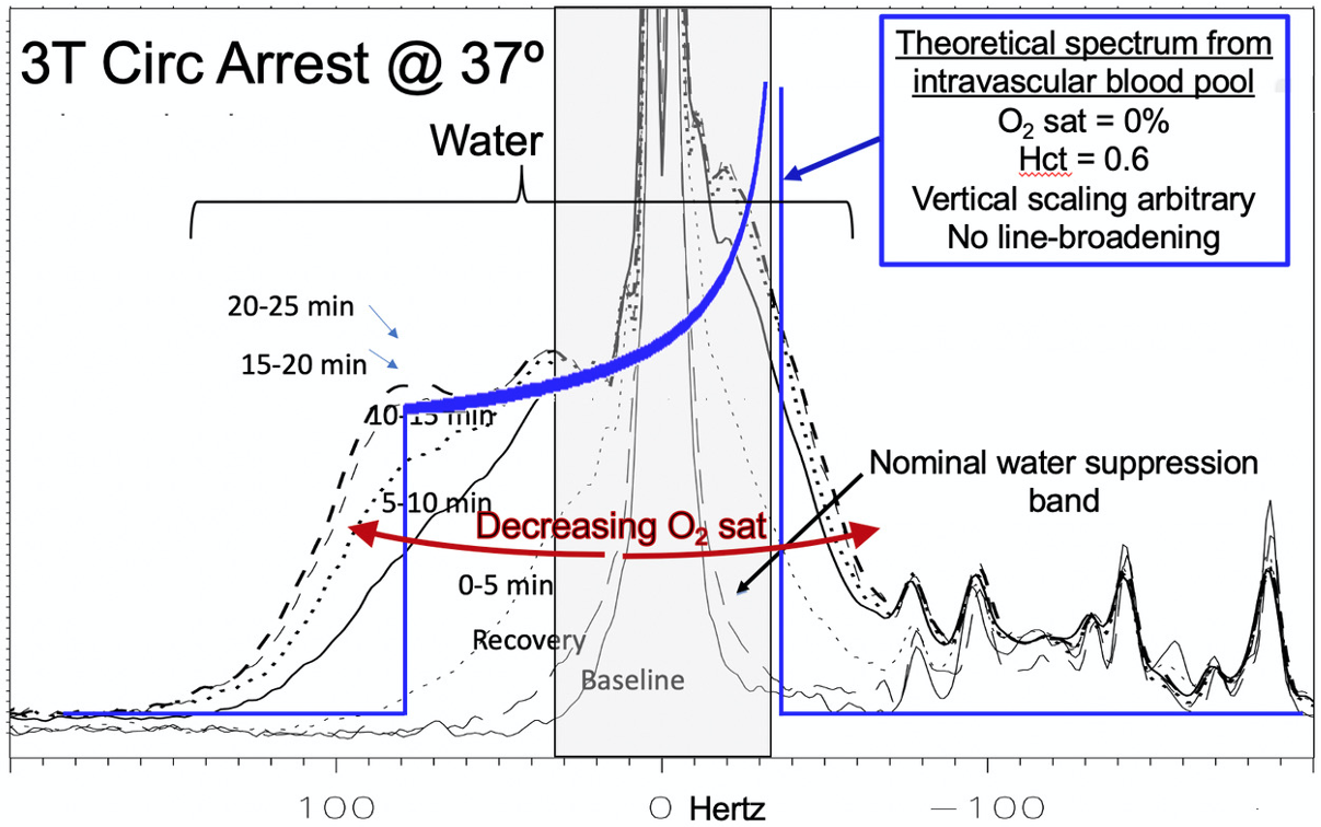
Figure 4. Observation of NMR “powder pattern” from residual water signal from a brain voxel in a neonatal pig model under circulatory arrest. Dynamic spectra are plotted as a function of time post-circulatory arrest. A powder pattern is theoretically predicted from the intravascular water signal, based on a model of randomly oriented veins/capillaries containing deoxyhemoglobin [5].