Parker L La1, Robyn Walker1, Julie M Joyce1, Tiffany Bell1, William Craig2, Quynh Doan3, Miriam Beauchamp4, Roger Zemek5, Pediatric Emergency Research Canada (PERC)6, Keith O Yeates7, and Ashley D Harris1
1Radiology, University of Calgary, Calgary, AB, Canada, 2Pediatrics, University of Alberta and Stollery Children's Hospital, Edmonton, AB, Canada, 3Pediatrics, University of British Columbia, Vancouver, BC, Canada, 4Psychology, University of Montreal and St Justine Hospital, Montreal, QC, Canada, 5Pediatrics and Emergency Medicine, University of Ottawa and Children's Hospital of Eastern Ontario, Ottawa, ON, Canada, 6Alberta Children's Hospital, Calgary, AB, Canada, 7Psychology, University of Calgary, Calgary, AB, Canada
1Radiology, University of Calgary, Calgary, AB, Canada, 2Pediatrics, University of Alberta and Stollery Children's Hospital, Edmonton, AB, Canada, 3Pediatrics, University of British Columbia, Vancouver, BC, Canada, 4Psychology, University of Montreal and St Justine Hospital, Montreal, QC, Canada, 5Pediatrics and Emergency Medicine, University of Ottawa and Children's Hospital of Eastern Ontario, Ottawa, ON, Canada, 6Alberta Children's Hospital, Calgary, AB, Canada, 7Psychology, University of Calgary, Calgary, AB, Canada
In the largest magnetic resonance spectroscopy dataset in pediatric concussion study to date, no differences in metabolites were found between concussion and control groups, and only Glx was related to cognitive symptoms.
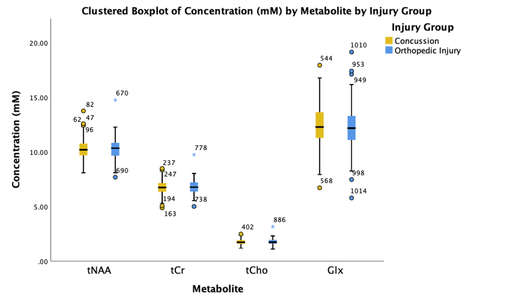
Figure 2: Boxplot showing the average concentration of tNAA, tCr, tCho, and Glx in both concussion and OI groups. There are no significant differences between concussion and OI groups in the aforementioned metabolites utilizing multiple ANCOVAs while controlling for age, sex, and site. Circles outside of the box plots are outside the 1.5 standard deviations interquartile range, and the stars are 3 standard deviations outside the interquartile range. All outliers meet all data quality criteria.
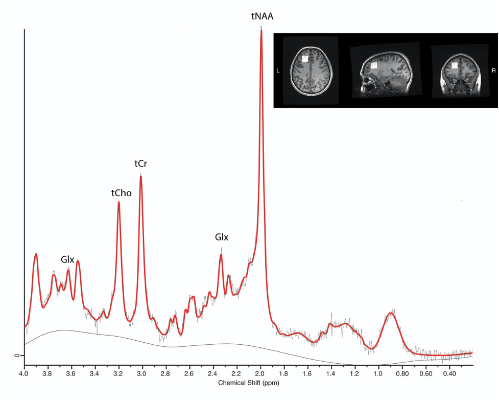
Figure 1: Example spectrum of a pediatric participant with concussion. This 1H-MRS PRESS data is from a 2x2x2 cm3 voxel in the left dorsolateral prefrontal cortex, shown in the voxel placements on the anatomical brain scan, to measure tNAA, tCr, tCho, and Glx. The PRESS sequence had a TE of 30ms and a TR of 2000ms, with 96 averages.