Alexandra Lipka1,2, Eva Heckova1, Assunta Dal-Bianco3, Gilbert Hangel1, Bernhard Strasser1, Stanislav Motyka1, Lukas Hingerl1, Paulus Rommer3, Fritz Leutmezer3, Petra Hnilicová4, Ema Kantorová4, Stephan Gruber1, Siegfried Trattnig1,2, and Wolfgang Bogner1,2
1High Field MR Centre, Department of Biomedical Imaging and Image-guided Therapy, Medical University of Vienna, Vienna, Austria, 2Christian Doppler Laboratory for Clinical Molecular MR Imaging, Vienna, Austria, 3Department of Neurology, Medical University of Vienna, Vienna, Austria, 4Jessenius Faculty of Medicine in Martin, Comenius University, Bratislava, Slovakia
1High Field MR Centre, Department of Biomedical Imaging and Image-guided Therapy, Medical University of Vienna, Vienna, Austria, 2Christian Doppler Laboratory for Clinical Molecular MR Imaging, Vienna, Austria, 3Department of Neurology, Medical University of Vienna, Vienna, Austria, 4Jessenius Faculty of Medicine in Martin, Comenius University, Bratislava, Slovakia
Free Induction Decay - Magnetic Resonance Spectroscopic Imaging (FID-MRSI) enables a comprehensive biochemical characterization of Multiple Sclerosis (MS) lesions and
strengthens the role of mIns/tNAA as an imaging biomarker for MS
pathologies.
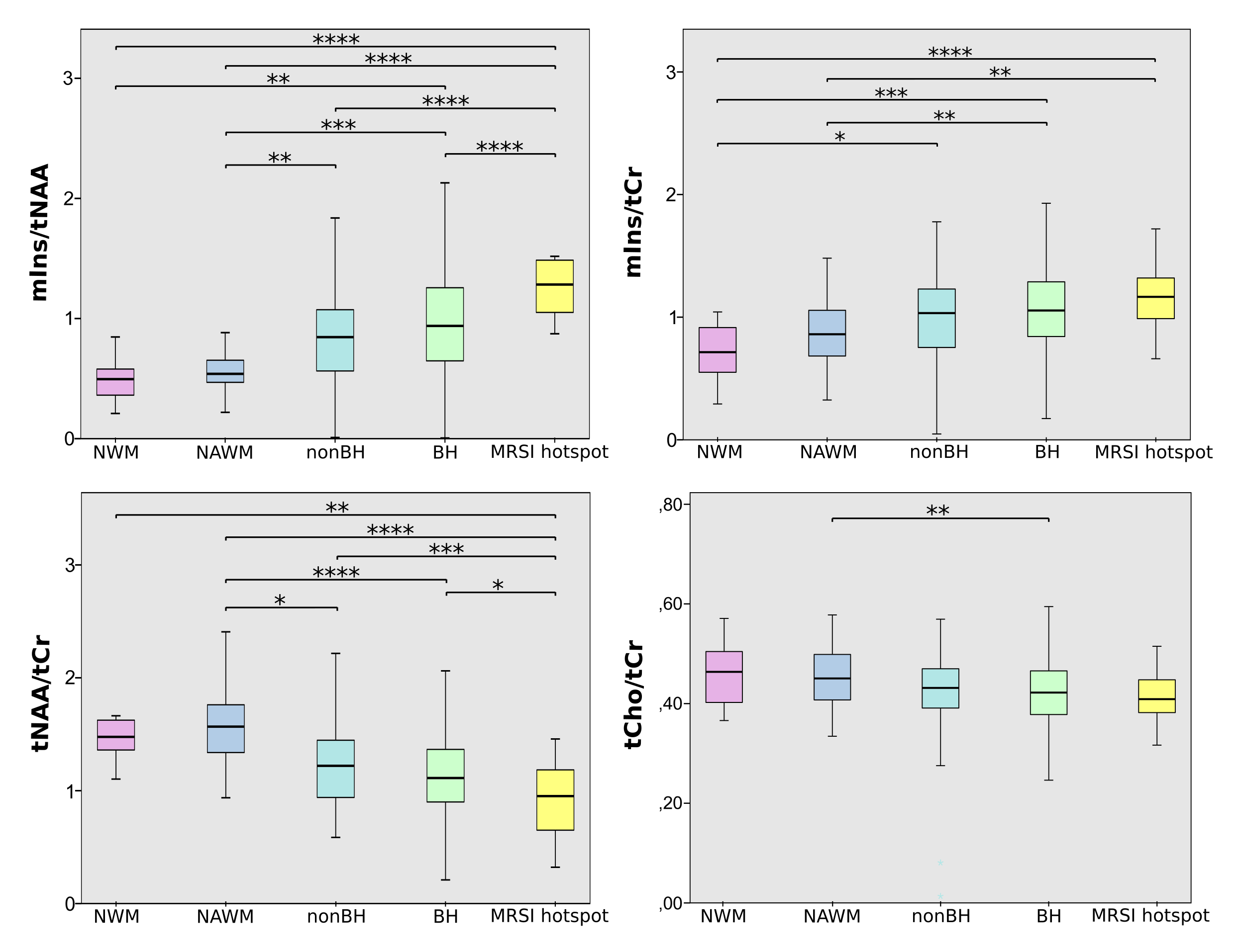
Figure 1: Boxplot
diagram for mIns/tNAA, mIns/tCr, tNAA/tCr and tCho/tCr and the
different lesion categories “NWM”, “NAWM”, “nonBH”, “BH”,
and “MRSI hotspot”. No differences between NWM and NAWM for each
of the metabolites. Highly significant differences between lesion
categories especially for mIns/tNAA
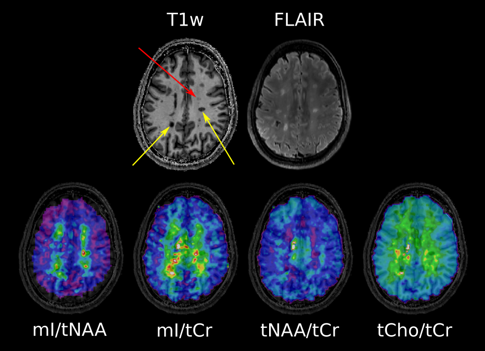
Figure 2:
Conventional MRI with T1-weighted MP2RAGE and T2-weighted FLAIR.
Yellow arrows point to “black hole” lesions which are clearly
visible on mIns/tNAA, mIns/tCr and tNAA/tCr. The red arrow depicts a
“non black hole” lesion, which is already apparent on mIns/tNAA
and mIns/tCr, though not yet visible on FLAIR