Moyoko Tomiyasu1,2, Jun Shibasaki3, Yasuhiko Terada4, Katsuaki Toyoshima3, Tatsuya Higashi1, Takayuki Obata1, and Noriko Aida2
1Department of Molecular Imaging and Theranostics, National Institute for Quantum and Radiological Science and Technology, Chiba, Japan, 2Department of Radiology, Kanagawa Children’s Medical Center, Yokohama, Japan, 3Department of Neonatology, Kanagawa Children’s Medical Center, Yokohama, Japan, 4Institute of Applied Physics, University of Tsukuba, Tsukuba, Japan
1Department of Molecular Imaging and Theranostics, National Institute for Quantum and Radiological Science and Technology, Chiba, Japan, 2Department of Radiology, Kanagawa Children’s Medical Center, Yokohama, Japan, 3Department of Neonatology, Kanagawa Children’s Medical Center, Yokohama, Japan, 4Institute of Applied Physics, University of Tsukuba, Tsukuba, Japan
The brain
temperature of neonates with hypoxic-ischemic encephalopathy was significantly
different during and after hypothermia, but was not significantly different
between favorable and adverse outcomes evaluated by neurodevelopmental testing at
18–22 months of age.
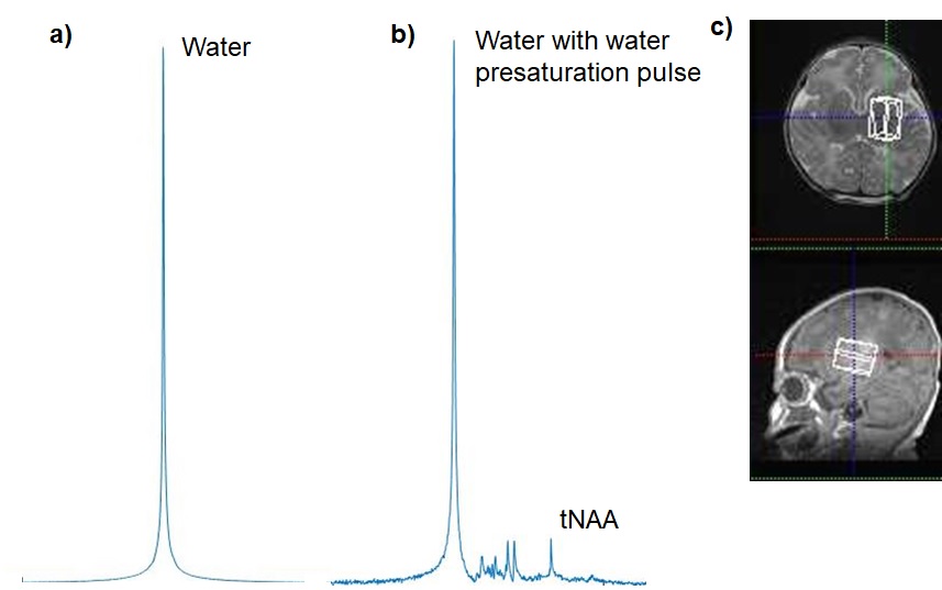
Figure 1. Representative in vivo proton MR spectra
for the deep gray matter obtained from a neonate with hypo-ischemic
encephalopathy: a) without and b) with water presaturation pulse in PRESS
sequence (TE/TR = 30/5000 ms). Scales of a) and b) are not proportional. c) White
voxel on the images show MR spectroscopy volumes of interest. tNAA =
N-acetylaspartate and N-acetylaspartylglutamate.
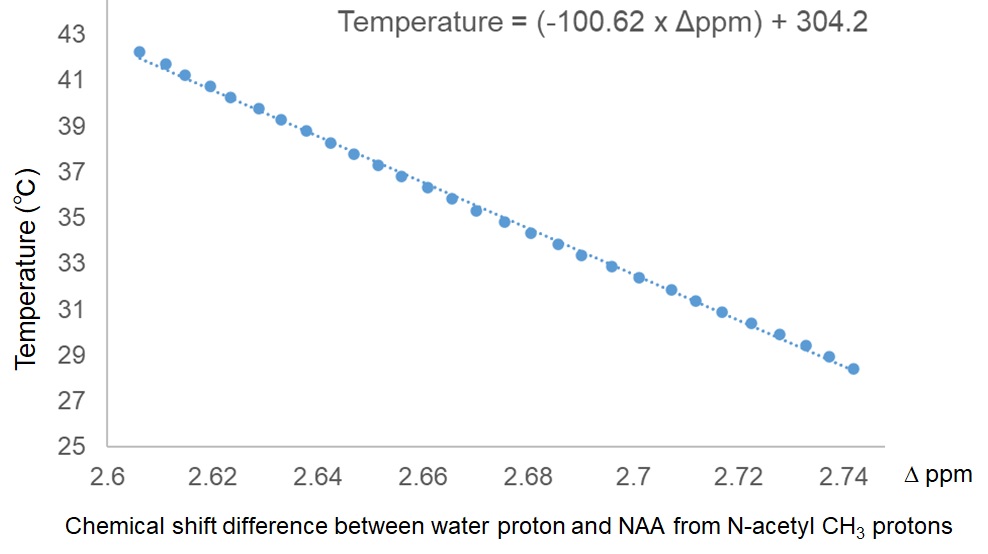
Figure
2. Calibration curve obtained from NAA aqueous solution.