Tingting Liu1, Weihao Zheng1, Yuqing You2, Ying Lv2, Weijun Chen2, Zhiyong Zhao1, Fusheng Gao2, Hongxi Zhang2, Chai Ji2, and Dan Wu1
1ZheJiang University, Hangzhou, China, 2Children's Hospital, ZheJiang University School of Medicine, Hangzhou, China
1ZheJiang University, Hangzhou, China, 2Children's Hospital, ZheJiang University School of Medicine, Hangzhou, China
Most
brain regions showed significant asymmetry and the asymmetry changed with brain
development. The MD-based LI showed an center-versus-peripheral pattern, and contrast
function lateralization with adult. Consistent leftward lateralization in white
matter was observed.
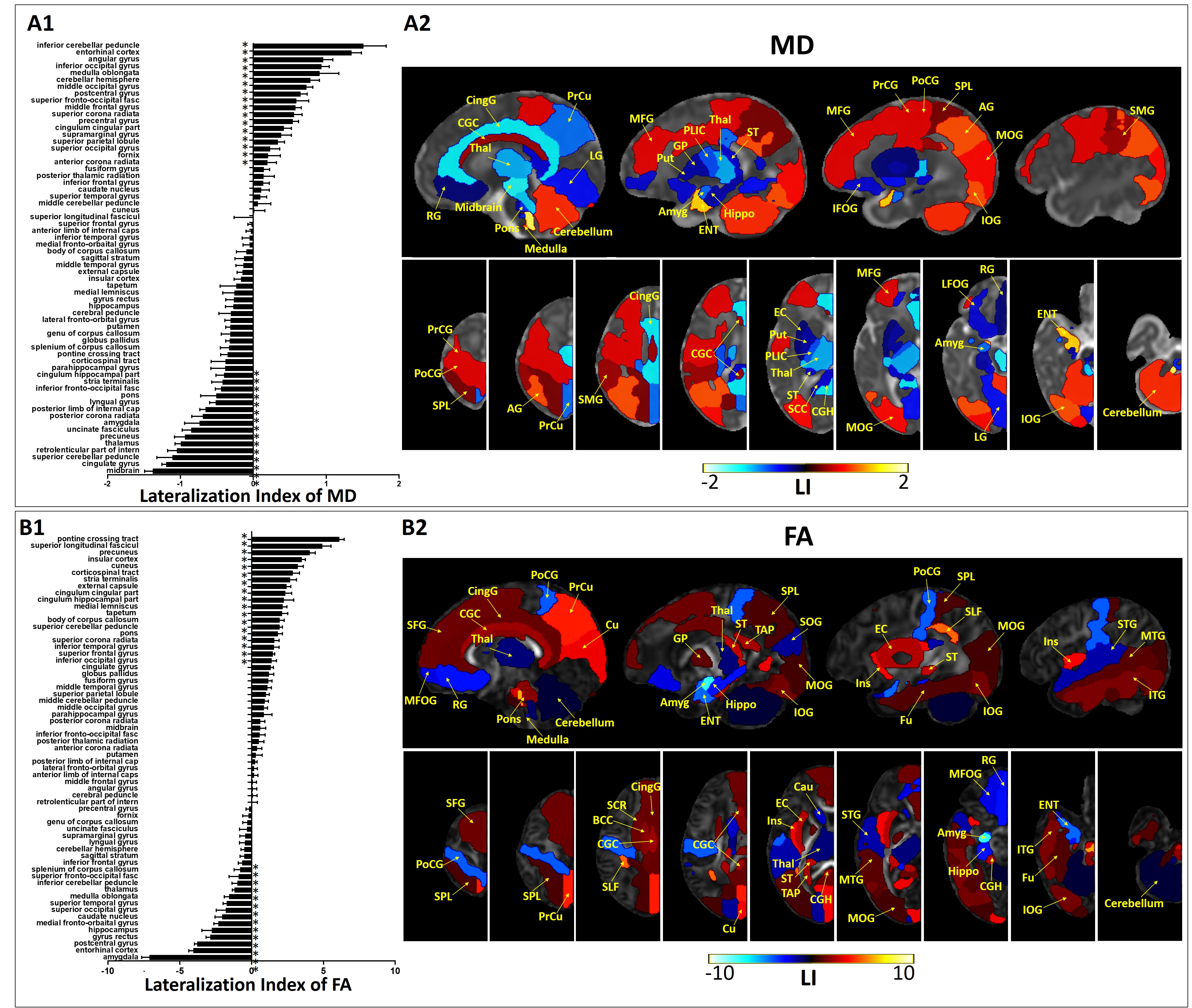
Figure 1.
Regions with significant left/right difference after multiple comparison
correction, based on MD (A) and FA (B). LIs of all 63 regions were plotted on
the left (A1 and B1), with the positive values indicating leftward asymmetry
and negative values indicating rightward asymmetry. *p < 0.05 by pairwise
t-test. Regions with significant asymmetry were overlaid on the corresponding
MD and FA maps in sagittal and axial views, with the color bar indicating the LI.
Positive values (red) represented leftward asymmetry, and negative values (blue)
represented rightward asymmetry.
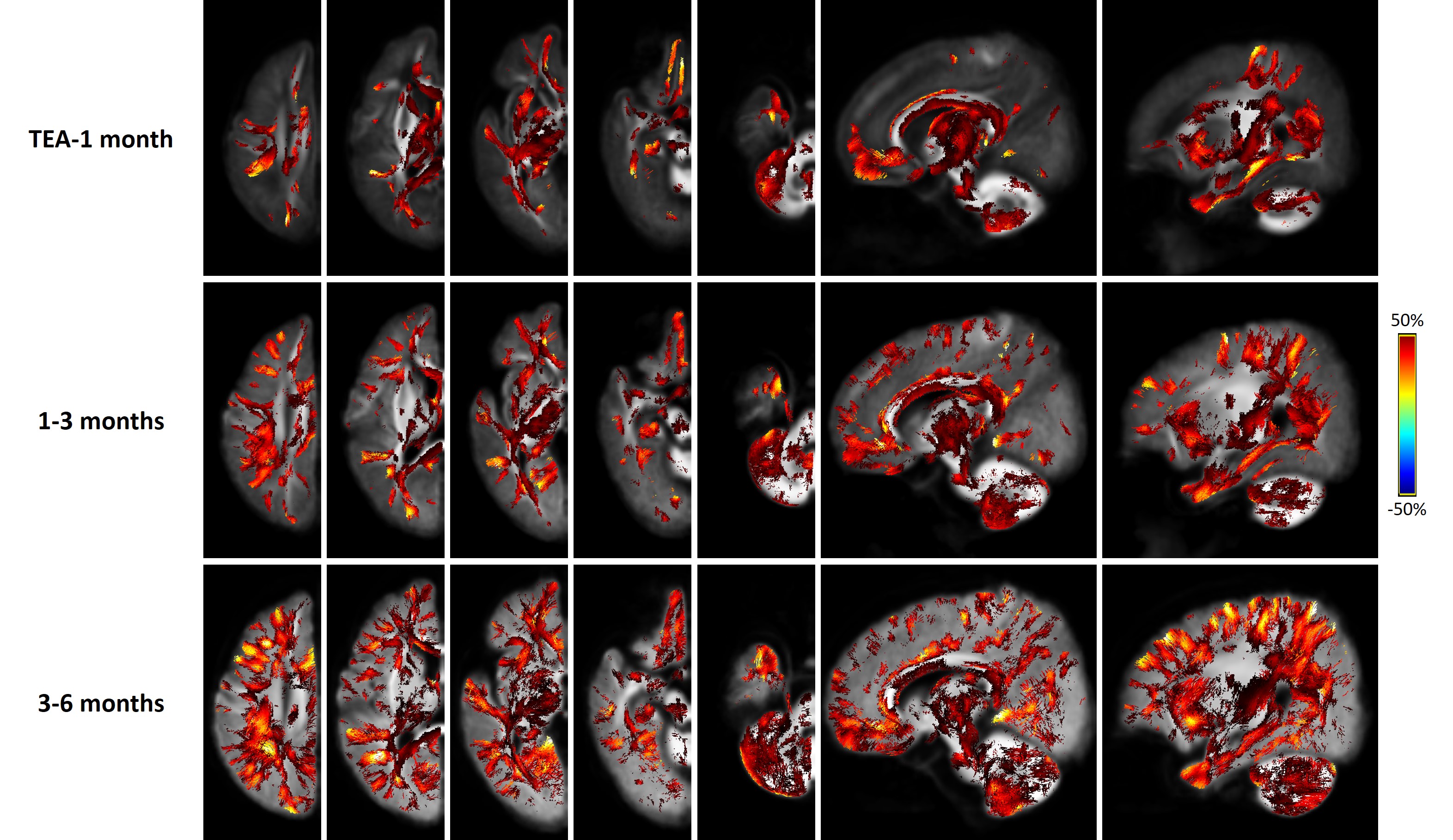
Figure 4.
FBA revealed extensive WM regions with significant asymmetry in terms of FDC.
The three rows from top to bottom show results for TEA-1month, 1-3 months, 3-6
months, respectively. The color bar indicates the LI, with positive values (red)
represent leftward asymmetry, and negative values (blue) represent rightward
asymmetry. A clear inside-to-outside developmental change can be
identified, with the lateralization primarily localizes in the central brain
and major WM at TEA, but extend to peripheral and subcortical WM at 1-3 months which
further increased at 3-6 months.