Wenchuan Wu1, Luke Baxter1,2, Eleri Adams2,3, Foteini Andritsou2, Matteo Bastiani1,4,5, Ria Evans Fry2, Robert Frost6,7, Sean Foxley8, Chris Gallagher1, Fiona Moultrie2, Vaneesha Monk2, David Andrew Porter9, Anthony Price10,11, Sebastian W Rieger1,12, Michael Sanders1, Anthony David Edwards11, Joseph V Hajnal10,11, Rebeccah Slater*1,2, and Karla L Miller*1
1Wellcome Centre for Integrative Neuroimaging, Nuffield Department of Clinical Neurosciences, University of Oxford, Oxford, United Kingdom, 2Department of Paediatrics, University of Oxford, Oxford, United Kingdom, 3Newborn Care Unit, John Radcliffe Hospital, Oxford University Hospitals NHS Foundation Trust, Oxford, United Kingdom, 4Sir Peter Mansfield Imaging Centre, School of Medicine, University of Nottingham, Nottingham, United Kingdom, 5NIHR Biomedical Research Centre, University of Nottingham, Nottingham, United Kingdom, 6Athinoula A. Martinos Center for Biomedical Imaging, Massachusetts General Hospital, Charlestown, MA, United States, 7Department of Radiology, Harvard Medical School, Boston, MA, United States, 8Department of Radiology, University of Chicago, Chicago, IL, United States, 9Imaging Centre of Excellence, College of Medical, Veterinary & Life Sciences, University of Glasgow, Glasgow, United Kingdom, 10Department of Biomedical Engineering, School of Biomedical Engineering and Imaging Sciences, King’s College London, King’s Health Partners, St. Thomas’ Hospital, London, United Kingdom, 11Centre for the Developing Brain, School of Biomedical Engineering and Imaging Sciences, King’s College London, King’s Health Partners, St. Thomas’ Hospital, London, United Kingdom, 12Department of Psychiatry, University of Oxford, Oxford, United Kingdom
1Wellcome Centre for Integrative Neuroimaging, Nuffield Department of Clinical Neurosciences, University of Oxford, Oxford, United Kingdom, 2Department of Paediatrics, University of Oxford, Oxford, United Kingdom, 3Newborn Care Unit, John Radcliffe Hospital, Oxford University Hospitals NHS Foundation Trust, Oxford, United Kingdom, 4Sir Peter Mansfield Imaging Centre, School of Medicine, University of Nottingham, Nottingham, United Kingdom, 5NIHR Biomedical Research Centre, University of Nottingham, Nottingham, United Kingdom, 6Athinoula A. Martinos Center for Biomedical Imaging, Massachusetts General Hospital, Charlestown, MA, United States, 7Department of Radiology, Harvard Medical School, Boston, MA, United States, 8Department of Radiology, University of Chicago, Chicago, IL, United States, 9Imaging Centre of Excellence, College of Medical, Veterinary & Life Sciences, University of Glasgow, Glasgow, United Kingdom, 10Department of Biomedical Engineering, School of Biomedical Engineering and Imaging Sciences, King’s College London, King’s Health Partners, St. Thomas’ Hospital, London, United Kingdom, 11Centre for the Developing Brain, School of Biomedical Engineering and Imaging Sciences, King’s College London, King’s Health Partners, St. Thomas’ Hospital, London, United Kingdom, 12Department of Psychiatry, University of Oxford, Oxford, United Kingdom
Results from our high-resolution post-mortem neonatal imaging platform demonstrate the ability to resolve features that are less prominent in low-resolution in-vivo data and potentially provide new anatomical insight into the developing brain.
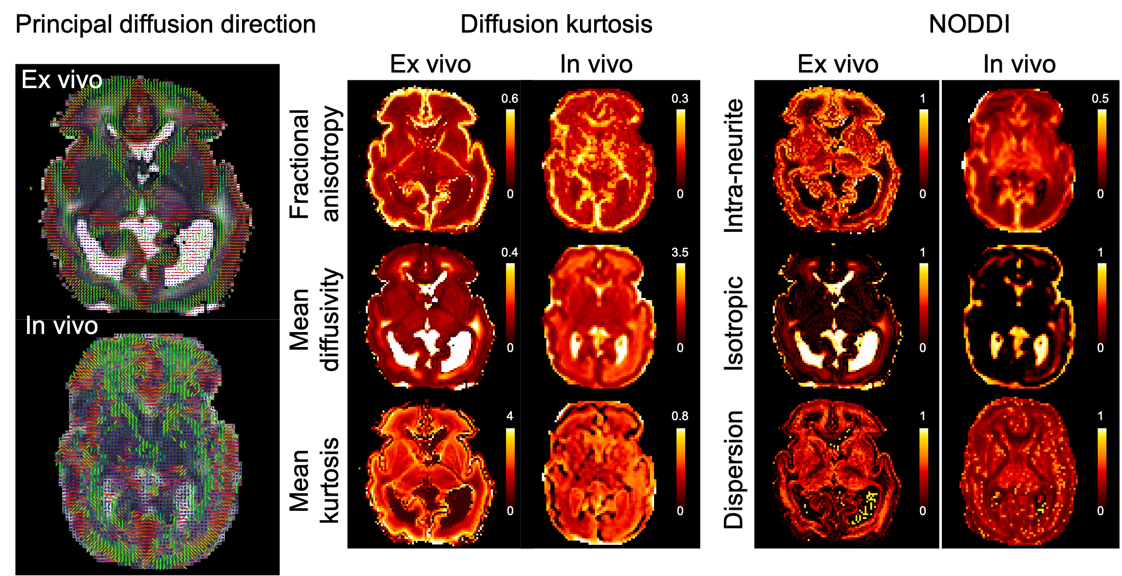
Figure 2. Diffusion kurtosis and NODDI parameters from the post-mortem infant and an age-matched in vivo infant. The mean diffusivity is expressed in μm2/ms.
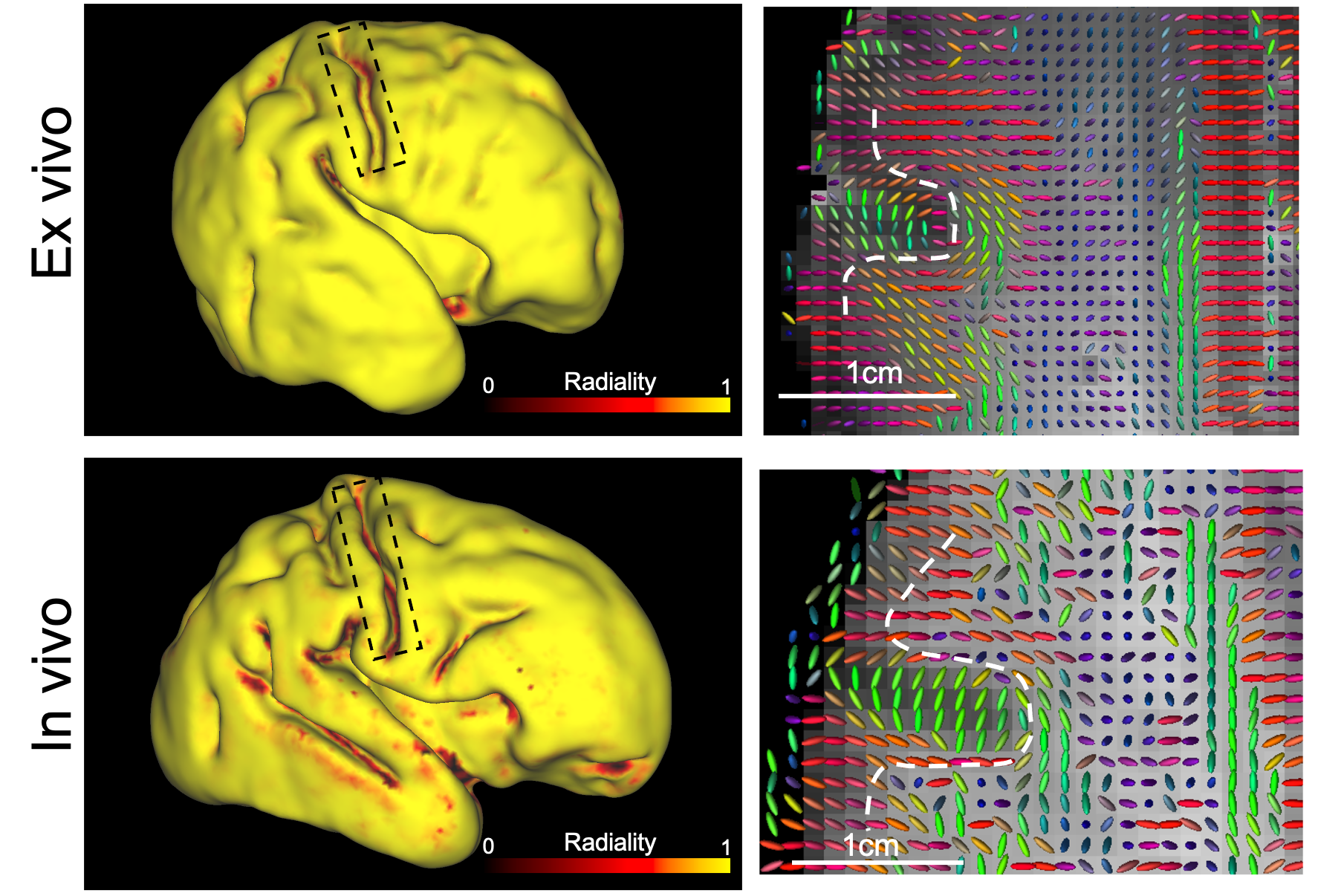
Figure 3. Left: Cortical radiality (dot product of principal diffusion direction and surface normal vector) of post mortem infant and an age-matched in vivo infant, calculated at mid-thickness. At the fundus of the central sulcus (black box), the post mortem data have high radiality but the in vivo data have low radiality. Right: principal diffusion directions for an axial plane containing central sulcus, the dashed white line indicates the grey white matter boundary. The principal diffusion directions at the fundus are radial in the post-mortem data but tangential in the in vivo data.
