Sila Genc1, Maxime Chamberland1, Gareth Ball2, Erika Raven1, Isobel Ward1, Chantal Tax1, Marco Palombo3, and Derek Jones1
1Cardiff University Brain Research Imaging Centre (CUBRIC), Cardiff University, Cardiff, United Kingdom, 2Developmental Imaging, Murdoch Children's Research Institute, Parkville, Australia, 3Centre for Medical Image Computing and Department of Computer Science, University College London, London, United Kingdom
1Cardiff University Brain Research Imaging Centre (CUBRIC), Cardiff University, Cardiff, United Kingdom, 2Developmental Imaging, Murdoch Children's Research Institute, Parkville, Australia, 3Centre for Medical Image Computing and Department of Computer Science, University College London, London, United Kingdom
In a sample of children and adolescents aged 8-18 years, we study developmental patterns of cortical microstructure using diffusion MRI. Our findings suggest an increase in neurite signal fraction and orientation dispersion with age, and a decrease in apparent soma radius with age.
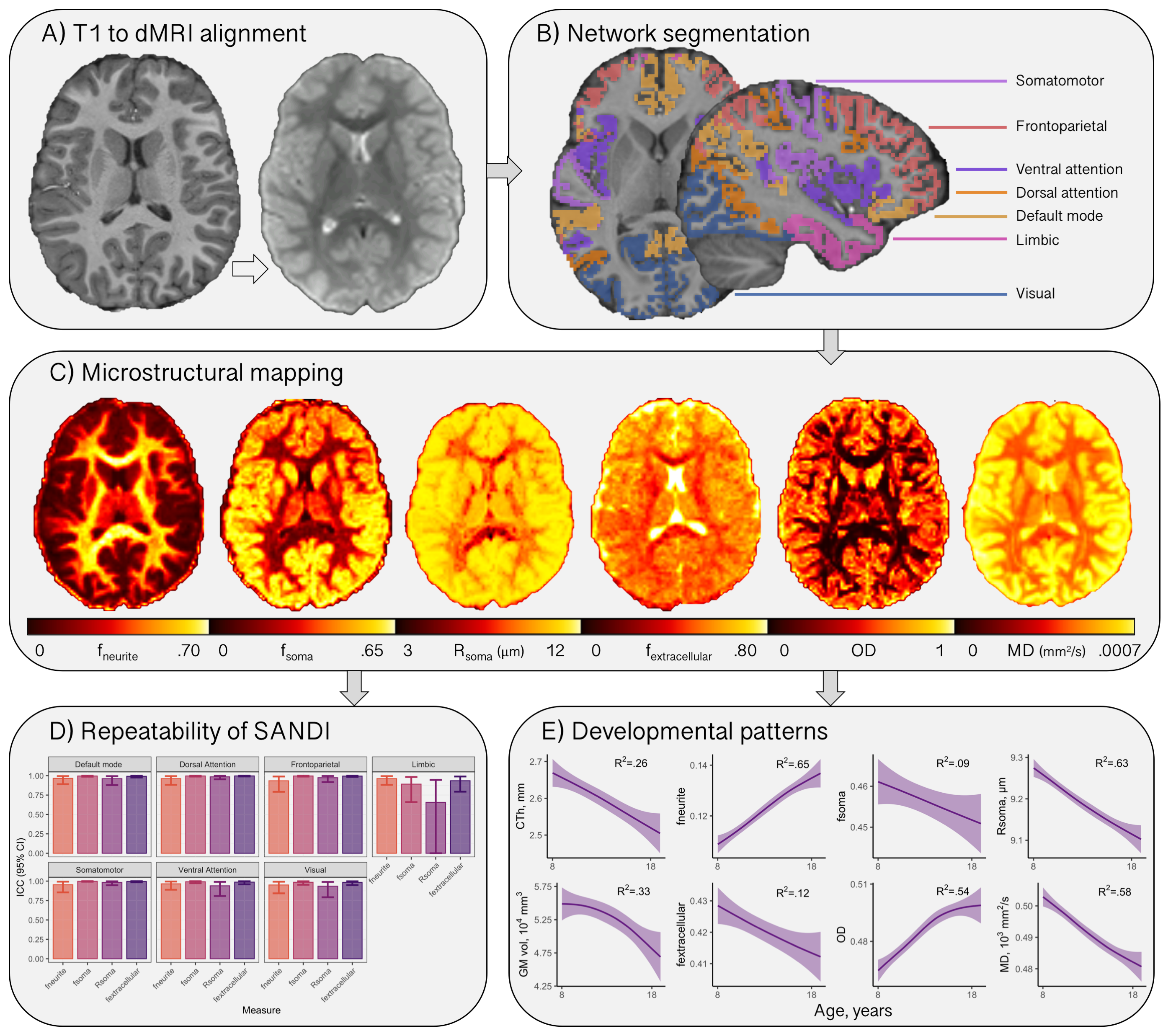
Figure 1: Cortical microstructure processing and analysis framework. A) T1-weighted data corrected for bias field and co-registered to an upsampled b=0 s/mm2 (1x1x1mm); B) T1-weighted data processed using Freesurfer[13,24], registered to MNI space to obtain 7 functionally defined networks[12], and resampled to dMRI space to obtain network labels intersecting the cortical ribbon; C) representative maps of microstructural measures; D) high repeatability of SANDI measures at 300mT/m[9]; E) developmental patterns of macro- and microstructure averaged over the cortical ribbon
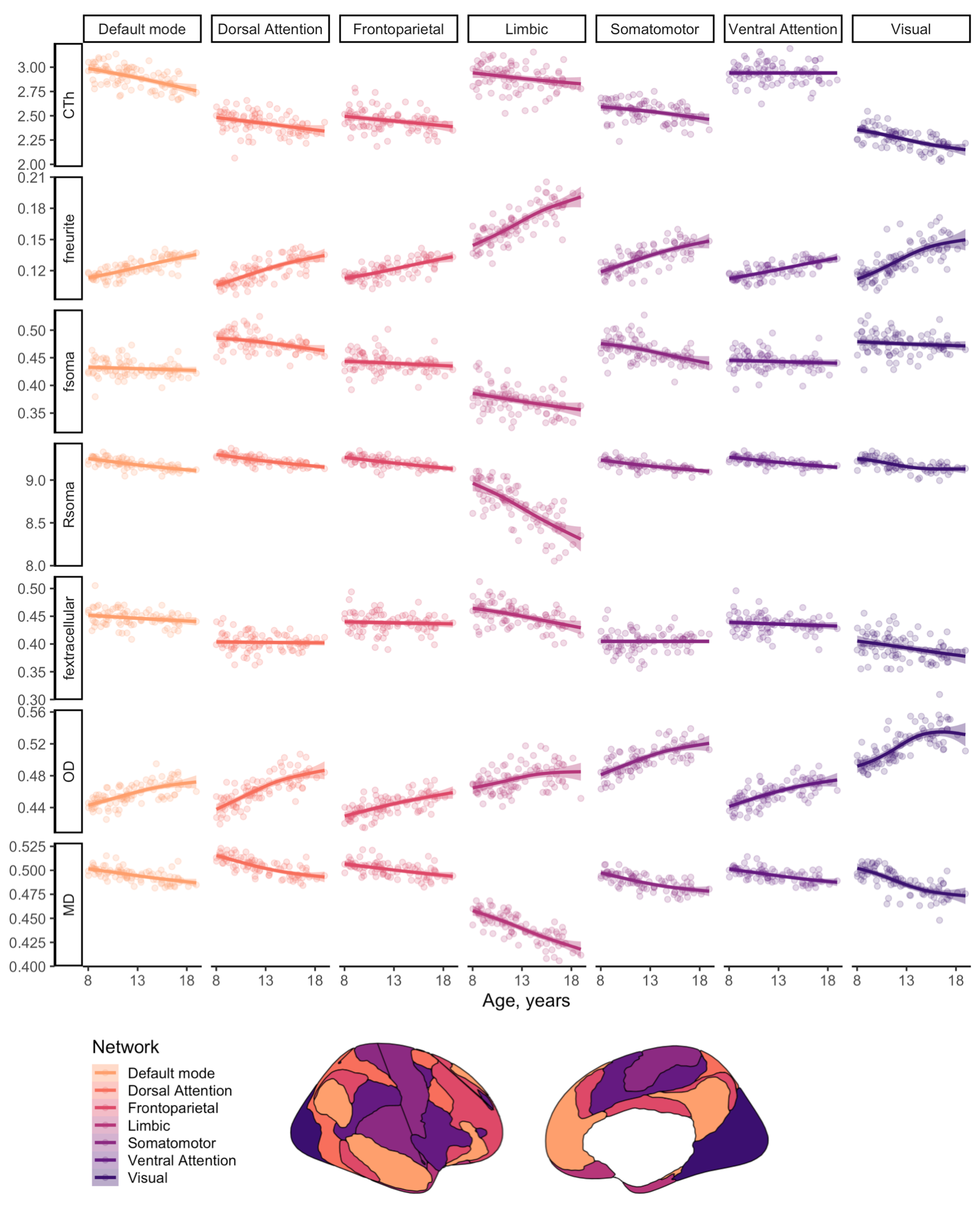
Figure 2: Relationship between cortical microstructure and age, grouped by measure and coloured by network type. Abbreviations: CTh = cortical thickness, in mm; f = signal fraction; MD = mean diffusivity, in 10-3 mm2/s; OD = orientation dispersion; R = apparent radius, in µm. Note: MD was estimated using b=0,6000 s/mm2 to improve sensitisation to cortical microarchitecture[17]