Sevgi Gokce Kafali1,2, Shu-Fu Shih1,2, Xinzhou Li1,2, Tess Armstrong1, Kelsey Kuwahara3, Sparsha Govardhan4, Karrie V Ly4, Shahnaz Ghahremani1, Kara L Calkins4, and Holden H Wu1,2
1Radiological Sciences, University of California, Los Angeles, Los Angeles, CA, United States, 2Bioengineering, University of California, Los Angeles, Los Angeles, CA, United States, 3Cognitive Science, University of California, Los Angeles, Los Angeles, CA, United States, 4Pediatrics, University of California, Los Angeles, Los Angeles, CA, United States
1Radiological Sciences, University of California, Los Angeles, Los Angeles, CA, United States, 2Bioengineering, University of California, Los Angeles, Los Angeles, CA, United States, 3Cognitive Science, University of California, Los Angeles, Los Angeles, CA, United States, 4Pediatrics, University of California, Los Angeles, Los Angeles, CA, United States
This work used a densely connected neural network with a class frequency balancing, boundary emphasizing loss to segment adipose tissue using free breathing MRI in children. The network achieved high Dice scores and similar volume and content quantification with reference annotations.

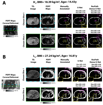
Figure 2. Representative images shown for (A) a healthy child (H1) and (B) a child with liver disease (L4). The coronal reformats show the chosen slices. The 2-D TE1 image and PDFF maps were inputs to the network. SAT (white) and VAT (gray) manual annotations are displayed. Both networks yielded comparable Dice-SAT. The proposed network yielded higher Dice-VAT than U-Net for H1 and L4, with differences shown in zoomed insets. The U-Net misclassified VAT as seen in violet boxes for H1 on chosen slice 2, and for L4 on chosen slices 1 and 2. Underlined Dice scores represent the higher score.

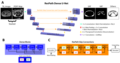
Figure 1. The structure of the proposed network. (A) ResPath Dense U-Net takes 2-D TE1 images and PDFF maps as inputs, and outputs segmentation masks of 3 classes: SAT, VAT, and the others. (B) Dense connections, shown inside a dense block, improve the generalizability of the features and overcomes the vanishing gradient problem. (C) ResPath skip connections mitigate the semantic gaps during the concatenation of encoder and decoder features.
