Amol Pednekar1, Deep B. Gandhi2, Hui Wang3, Jean A. Tkach1, Andrew T. Trout1, and Jonathan R. Dillman1
1Department of Radiology, Cincinnati Children's Hospital Medical Center, Cincinnati, OH, United States, 2Imaging Research Center, Department of Radiology, Cincinnati Children's Hospital Medical Center, Cincinnati, OH, United States, 3MR Clinical Science, Philips, Cincinnati, OH, United States
1Department of Radiology, Cincinnati Children's Hospital Medical Center, Cincinnati, OH, United States, 2Imaging Research Center, Department of Radiology, Cincinnati Children's Hospital Medical Center, Cincinnati, OH, United States, 3MR Clinical Science, Philips, Cincinnati, OH, United States
In 19 participants, mean liver shear stiffness
values measured with SC_2D_4BH and 2D_CS_HBH with or without flow saturation
correlated very strongly (ICC>0.96) with mean bias of <0.15 kPa (<6 %). 2D_CS_HBH MRE has potential
benefit in participants with compromised breath-holding capacity.
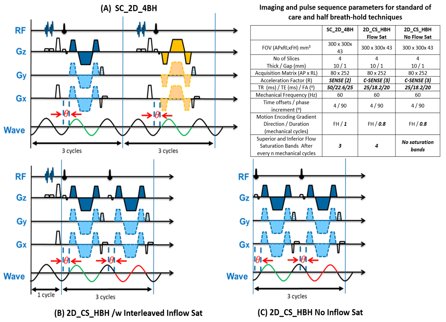
Figure 1: Schematic pulse sequence diagrams. (A) SC_2D_4BH: The polarity of MEGs is
reversed (blue and yellow) every RF excitation. The mechanical wave polarity
(green) stays the same across each RF excitation. Each RF excitation triggers 3
motion cycles. (B) 2D_CS_HBH with inflow saturation: The polarity of MEGs
remains the same (blue) across RF excitations. The mechanical wave polarity
inverts (green and red) every RF excitation. (C) 2D_CS_HBH no inflow
saturation. Every other RF excitation triggers 4/3 motion cycles.
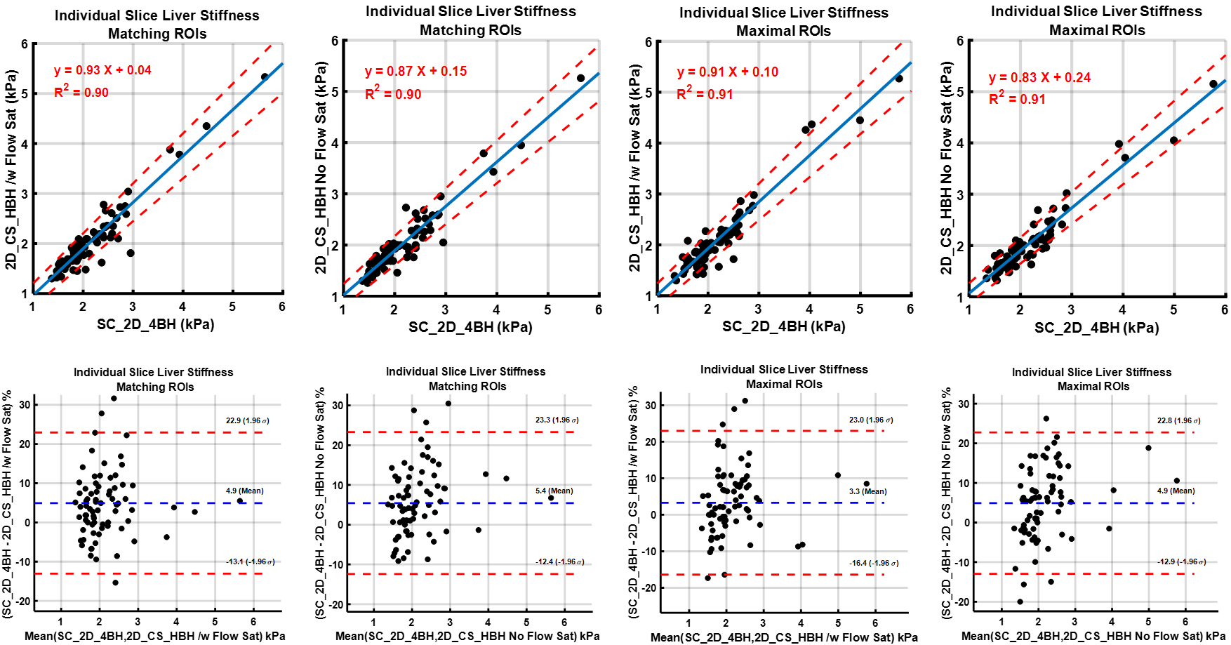
Figure 3: Comparison of liver shear stiffness values
in individual slices for SC_2D_4BH and 2D_CS_HBH techniques by Linear Regression and Bland-Altman Analysis. Values
are based on manual analysis informed by a 95% confidence mask with matching
ROIs on images from both techniques and maximal possible ROI in each. SC_2D_4BH:
standard of care two-dimensional 4 slices through mid-liver with 13 second
breath-hold per slice; 2D_CS_HBH: two-dimensional polarity-inversion motion encoding plus compressed SENSE acquisition in half the SC breath-hold.