Diego M Jaramillo1, Phuong M Duong1, Jie C Nguyen2, Sogol Mostoufi-Moab2, Michael K Nguyen2, Andrew Moreau2, Christian A Barrera3, Shijie Hong2, and Jose M Raya4
1Columbia University Medical Center, New York, NY, United States, 2Children’s Hospital of Philadelphia, Philadelphia, PA, United States, 3Massachusetts General Hospital, Boston, MA, United States, 4New York University, New York, NY, United States
1Columbia University Medical Center, New York, NY, United States, 2Children’s Hospital of Philadelphia, Philadelphia, PA, United States, 3Massachusetts General Hospital, Boston, MA, United States, 4New York University, New York, NY, United States
DTI of the physis and metaphysis (DTI-P/M) has potential to become a biomarker of skeletal growth. Compared to clinical standards DTI-P/M reduced prediction errors over 40% and has not bias in their predictions.
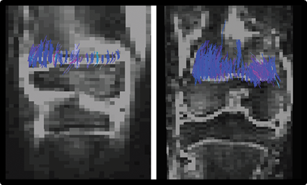
Figure 1. Example of tractography. Example of tractography of two children, ages 12 (boy) and 15 (girl).
Figure
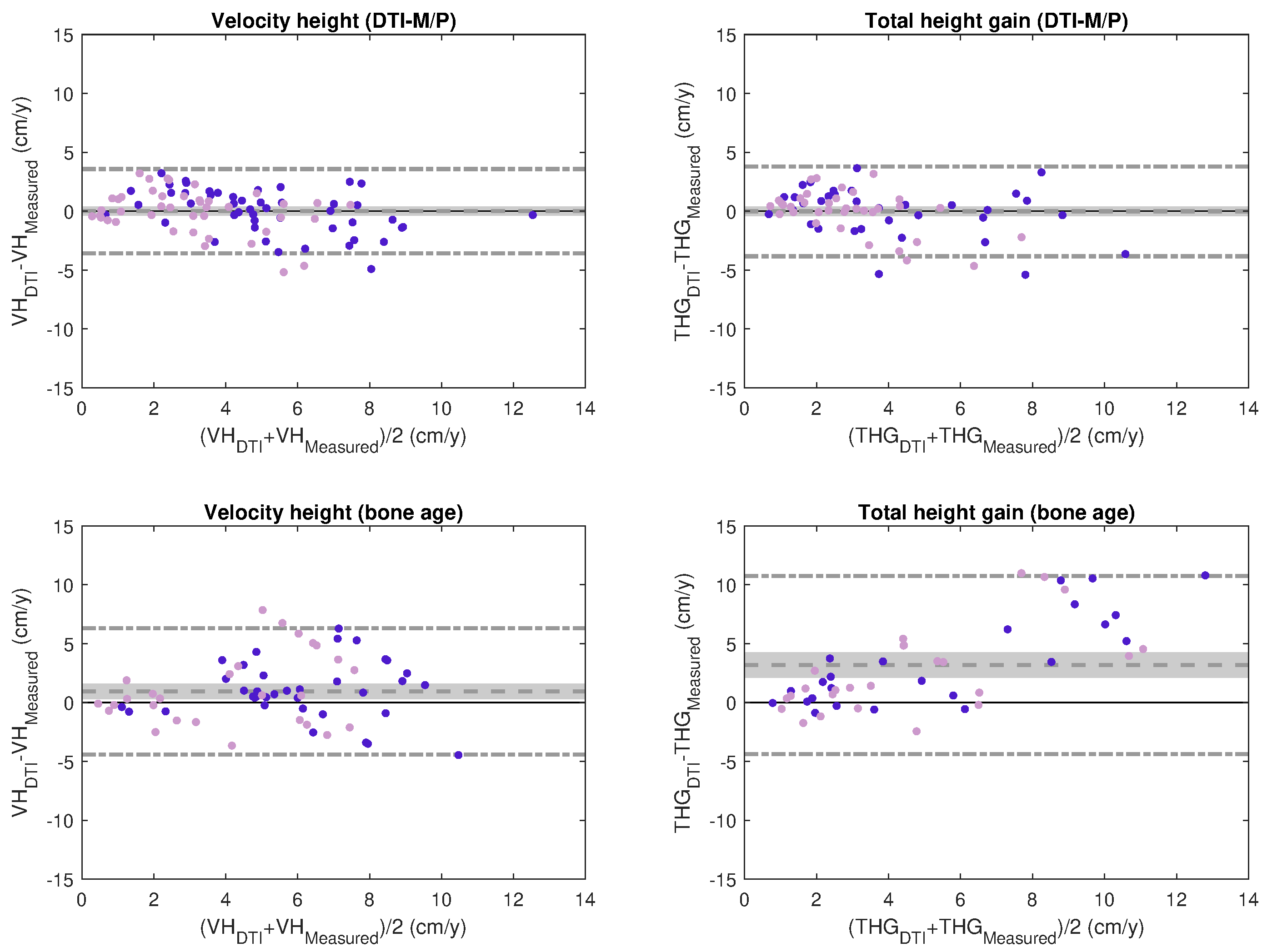
1. Bland-Altman plots of predicted and measured VH and THG. Bland-Altman plots
showed reduced error in DTI-P/M predictions (top) compared to bone-age
predictions (bottom). Also, DTI-P/M predictions did not have bias (zero
difference within the 95%-confidence interval), while bone-age predictions had
a significant bias. The dashed line represents the mean difference between
predicted and measured; the dot-dash line the two-sigma range. The light gray
area indicates the 95%-confidence interval of the mean difference. Purple
circles are boys and dark pink girls.