Xianyun Cai1, Jinxia Zhu2, and Guangbin Wang1
1Shandong Medical Imaging Research Institute, Shandong University, Jinan, China, China, 2MR Collaboration, Siemens Healthcare Ltd., Beijing, China, China
1Shandong Medical Imaging Research Institute, Shandong University, Jinan, China, China, 2MR Collaboration, Siemens Healthcare Ltd., Beijing, China, China
This study investigated the prenatal diagnosis and prognosis of fetal sacrococcygeal teratomas (SCTs) (Type IV) with Prenatal MRI. This suggests correct diagnosis of teratoma on prenatal MRI and early operation after birth are important for prognosis.

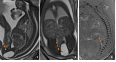
Fig 1. 25-weeks’ gestation fetus
with sacrococcygeal type IV teratoma
(a) (b) Sagittal and coronal
T2-weighted image shows cystic mass (arrow) arising from coccyx, with septa
(small arrow) is seen (b).
(c) Sagittal
Susceptibility-weighted imaging (SWI) shows
fetal sacrococcygeal vertebra
were nicely demonstrated

Fig 4. 28-weeks’ gestation fetus with sacrococcygeal type IV teratoma(a) (b) Coronal and sagittal T2-weighted image shows solid mass (arrow) arising from sacrococcygeal region with unilateral hydronephrosis (white arrow)(c,d) Diffusion Weighted Imaging (DWI) and corresponding apparent diffusion coefficient (ADC) showed the mass diffusion limited
