Lydia Jean Bardwell Speltz1,2, Yunhong Shu2, Myung-Ho In2, Nolan Meyer1,2, Erin Gray2, Diana Lanners2, Yihe Hua3, Robert E Watson2, John Huston III2, Thomas KF Foo3, and Matt A Bernstein2
1Mayo Clinic Graduate School of Biomedical Sciences, Mayo Clinic, Rochester, MN, United States, 2Department of Radiology, Mayo Clinic, Rochester, MN, United States, 3GE Global Research, Niskayuna, NY, United States
1Mayo Clinic Graduate School of Biomedical Sciences, Mayo Clinic, Rochester, MN, United States, 2Department of Radiology, Mayo Clinic, Rochester, MN, United States, 3GE Global Research, Niskayuna, NY, United States
A software tool reports B0, gradient slew rate, B1+2,
and dB/dz at a device location to assess if MR conditional devices can be
scanned safely on the compact 3T scanner.
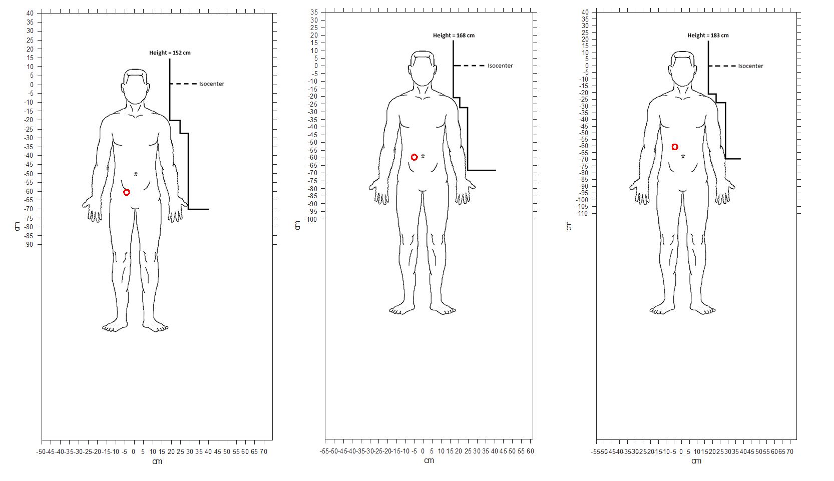
Figure 1: Senza IPG location based on three different heights indicated
as a red circle.
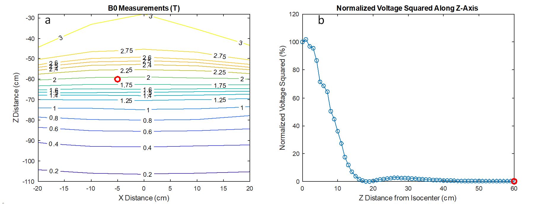
Figure 2: For
the specified location of the device (red circle), shown are a) the main
magnetic field strength and b) normalized RF voltage squared along the z-axis,
which is a measure of (B1+)2. Because the RF is negligible at the
location of the device, even if the device labeling calls for a T/R head coil, the
brain could be scanned with a multi-channel receive head coil, which offers a
substantial improvement in image quality.