Yingyu Lin1, Mengzhu Wang2, and Shi-Ting Feng1
1The First Affiliated Hospital, Sun Yat-sen University, Guangzhou, China, 2MR Scientific Marketing, Siemens Healthcare, Guangzhou, China
1The First Affiliated Hospital, Sun Yat-sen University, Guangzhou, China, 2MR Scientific Marketing, Siemens Healthcare, Guangzhou, China
We
compared MRI with EUS in preoperative T staging and with PET/CT in preoperative
lymphatic metastases of esophageal cancer. In this study, MRI showed higher
sensitivity, specificity and accuracy in T staging, while MRI and PET/CT showed similar performance in lymph nodes evaluation.
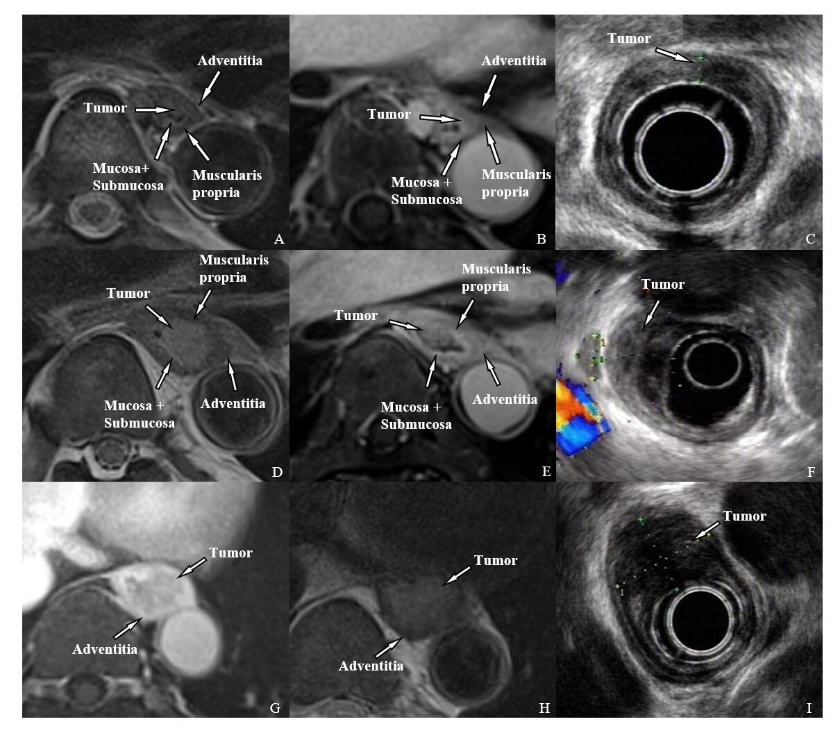
Figure1. MRI and EUS images of T1-T3 esophageal cancer. T1: A-C, T2WI BLADE(A), Enhanced T1WI radial VIBE(B), and EUS(C). The tumor located in the mucosa and submucosa and protrudes into the lumen without muscularis propria invasion. T2: D-F, T2WI BLADE(D), Enhanced T1WI radial VIBE(E), and EUS(F). The tumor breaks through the submucosa and is limited to the muscularis propria. T3:G-I, T2WI BLADE(G), Enhanced T1WI radial VIBE(H), and EUS(I). The tumor is confined to the adventitia.
Table
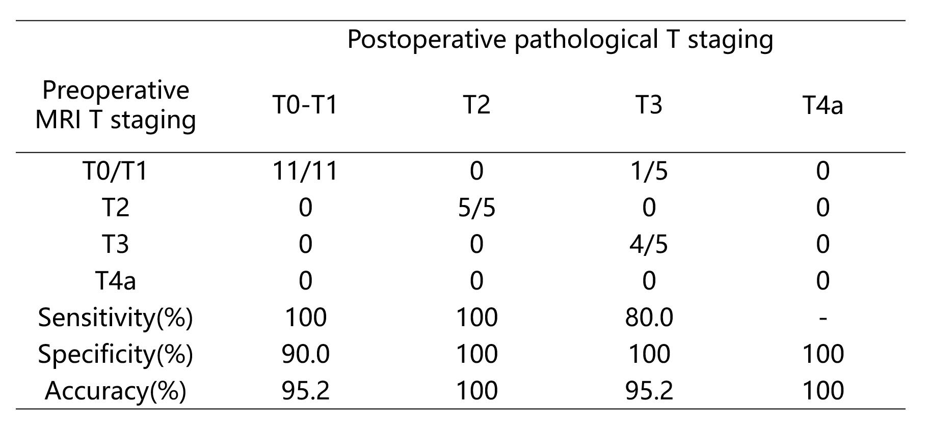
1. Comparison between preoperative MRI and postoperative pathological T staging