Deshuo Dong1, Ailian liu1, Jiazheng Wang2, Peng Sun2, Anliang Chen1, Wan Dong1, Yuhui Liu1, Qingwei Song1, and Renwang Pu1
1Radiology, The First Affiliated Hospital of Dalian Medical University, Dalian, China, 2Philips Healthcare, Beijing, China
1Radiology, The First Affiliated Hospital of Dalian Medical University, Dalian, China, 2Philips Healthcare, Beijing, China
This
study showed that T2 mapping combined with DKI had a better performance in the differential
diagnosis of rectal
cancer with and without vascular invasion.
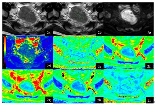
Figure 2. A 65-year-old
male rectal cancer patient without vascular invasion. T2W (1a), T2W with
ROI s(1b), DWI (1c), T2 mapping (1d) , FA (1e) , MD (1f), Da (1g), Dr (1h) , MK
(1i) map. Three ROIs of rectal cancer were showed on T2W image. Average T2, FA,
MD, Da , Dr, MK values of the three ROI were 83.85ms, 0.251, 0.705um2
ms−1, 0.725 um2 ms−1, 0.612
um2 ms−1, 0.419.
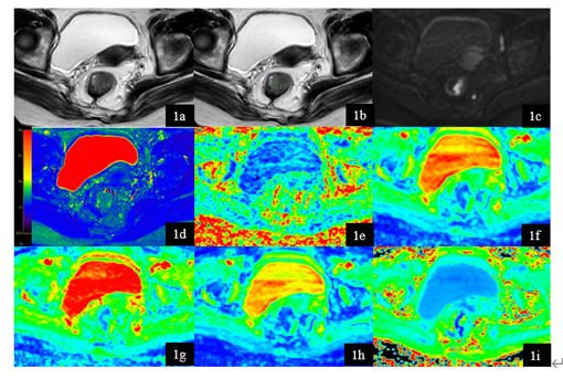
Figure 1. A 74-year-old
female rectal cancer patient with vascular invasion. T2W (1a), T2W with
ROI s(1b), DWI (1c), T2 mapping (1d) , FA (1e) , MD (1f), Da (1g), Dr (1h) , MK
(1i) map. Three ROIs of rectal cancer were showed on T2W image. Average T2, FA,
MD, Da , Dr , MK values of the three ROI were 80.47ms, 0.302, 1.013 um2
ms−1, 1.313 um2 ms−1, 0.866
um2 ms−1, 2.400.