Changjun Ma1, Ailian Liu1, Dahua Cui1, Ying Zhao1, Hongkai Wang2, and Mingrui Zhuang2
1Radiology Department, The First Affiliated Hospital of Dalian Medical University, Dalian, China, Dalian,China, China, 2the School of Biomedical Engineering, Dalian University of Technology, Dalian,China, China
1Radiology Department, The First Affiliated Hospital of Dalian Medical University, Dalian, China, Dalian,China, China, 2the School of Biomedical Engineering, Dalian University of Technology, Dalian,China, China
The aim of this study was to explore the value of intratumoral susceptibility signal intensities (ITSS) in quantitatively and automatically differentiating intrahepatic cholangiocarcinoma (ICC) from hepatocellular carcinoma (HCC) using enhanced T2 star-weighted angiography (ESWAN).
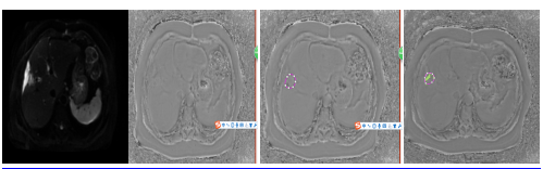
Figure2:A 66 year-old male withICC in the right lobe of the liver. (a)DKI image; (b) phase map; (c) tumor was delineated around the edge of the tumor; (d) AS software recognized quantitatively and automatically ITSS ratio by reading phase maps. ITSS recognized were covered in green.
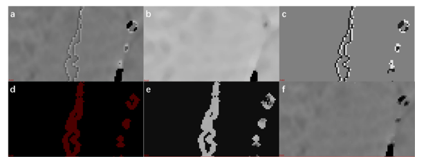
Figure1 workflow. (a) Input phase map that needs to be de-artifacted. (b) The median filtering result of A. (c) A and B are subtracted and threshold processed to obtain high pixel value and low pixel value bands. (d) The adjacent regions of high pixel value and low pixel value are extracted and expanded in the acquisition plane. This result is considered as the artifact region. (e) The result of recomputing the pixel values in the artifact region. The method is to calculate the average value of 26 adjacent non-artifact region pixels of each target pixel. (f) The result of removing the artifact.