Ting Jiang1, Diego Hernando2, Scott B. Reeder2, and Jin Wang1
1Radiology, The Third Affiliated Hospital of Sun Yat-Sen University, Guangzhou, China, 2University of Wisconsin, Madison, WI, United States
1Radiology, The Third Affiliated Hospital of Sun Yat-Sen University, Guangzhou, China, 2University of Wisconsin, Madison, WI, United States
Our
study suggests that high signal distribution around the tumor on R2* can predict MVI of HCC. We hypothesize that arterial-portal
shunting caused by MVI may explain abnormal peritumoral iron deposition.
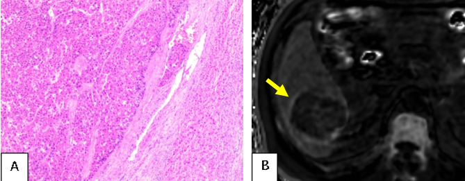
Figure 1. Surgically confirmed moderately differentiated HCC in a 59-year-old man with microvascular invasion found at pathology. HCC with microvascular invasion (hematoxylin-eosin stain, Original magnification 200×) (A). R2* map shows a hepatic mass(59mm×61mm) in S6 with elevated R2* around the tumor (B).
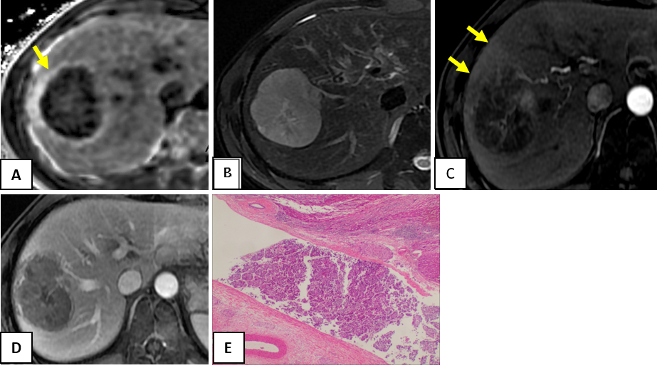
Figure 2. Surgically confirmed poorly differentiated HCC in a 55-year-old man with microvascular invasion found at pathology. R2* of CSE-MRI shows a hepatic mass(75mm×81mm) in S7/8 with elevated R2* around the tumor (A). The tumors in S7/8 show high signal intensity on FS -T2WI (B).The tumors in S7/8 show APHE and peritumoral corona enhancement and on early arterial phase and washout on the portal vein phase(C and D). HCC with microvascular invasion (hematoxylin-eosin stain, Original magnification 200×) (E).