Guangzi Shi1, Hong Chen1, Weike Zeng1, Mengzhu Wang2, and Jun Shen1
1Radiology, Sun Yat-Sen Memorial Hospital, Sun Yat-Sen University, Guangzhou, China, 2Department of MR Scientific Marketing, Siemens Healthineers, Guangzhou, China
1Radiology, Sun Yat-Sen Memorial Hospital, Sun Yat-Sen University, Guangzhou, China, 2Department of MR Scientific Marketing, Siemens Healthineers, Guangzhou, China
R2* value of malignant FLLs was significantly higher than that of the benign
FLLs. R2* derived from multi-echo Dixon imaging is a potential biomarker to
differentiate malignant from benign FFLs.
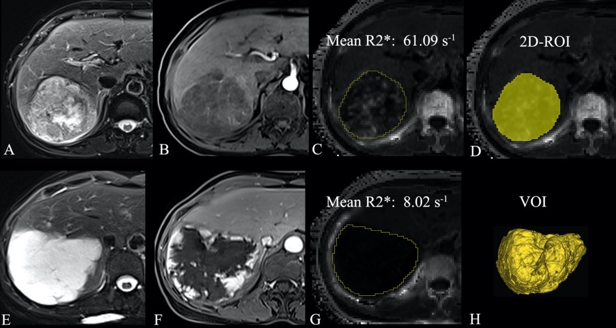
Figure
1. 2D region of
interest (2D-ROI) and volume of interest (VOI). T2-weighted imaging (T2WI) (A)
and arterial phase contrast-enhanced T1-weighted imaging (T1WI) (B), and R2*
map (C) show liver metastasis (yellow line) confirmed by histology in a
59-year-old woman with lung cancer. T2WI (E) and arterial phase
contrast-enhanced T1WI (F), and R2* map (G) show a live hemangioma (yellow
line) in in a 59-year-old woman. (D) 2D-ROI was drawn on the section showing
the maximal tumor dimension. (H) VOI was placed covering the
entire tumor volume on R2* map.
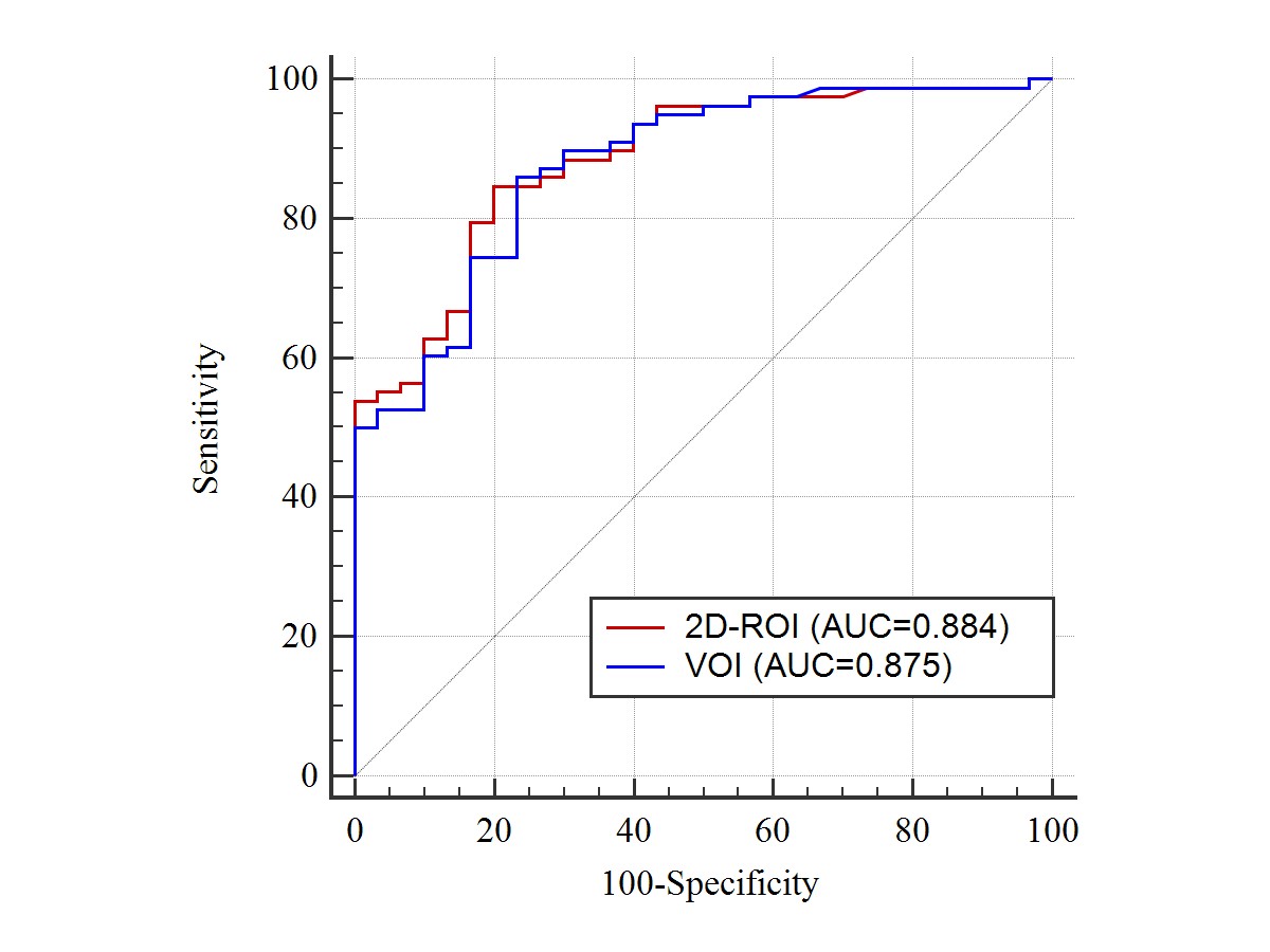
Figure
4. Receiver operating characteristic curve analysis of
the two positioning methods in differentiating
between malignant group and benign group. 2D region of interest (2D-ROI) and
volume of interest (VOI) methods yielded the similar results.