Puneet Bagga1, Jeffrey Steinberg2, Walter Akers2, Matthew Scoggins1, Zoltan Patay1, Beth McCarville1, Burkhard Hoeckendorf3, Khaled Khairy3, Michael Dyer3, and Beth Stewart3
1Department of Diagnostic Imaging, St Jude Children's Research Hospital, Memphis, TN, United States, 2Center for In Vivo Imaging and Therapeutics (CIVIT), St Jude Children's Research Hospital, Memphis, TN, United States, 3Department of Developmental Neurobiology, St Jude Children's Research Hospital, Memphis, TN, United States
1Department of Diagnostic Imaging, St Jude Children's Research Hospital, Memphis, TN, United States, 2Center for In Vivo Imaging and Therapeutics (CIVIT), St Jude Children's Research Hospital, Memphis, TN, United States, 3Department of Developmental Neurobiology, St Jude Children's Research Hospital, Memphis, TN, United States
In this study, we used multi-modal MRI/MRS methods including T2-weighted MRI, diffusion-weighted MRI, and 1H MRS in a PDX model of EWS to evaluate the treatment response and predict treatment response in the tumors.
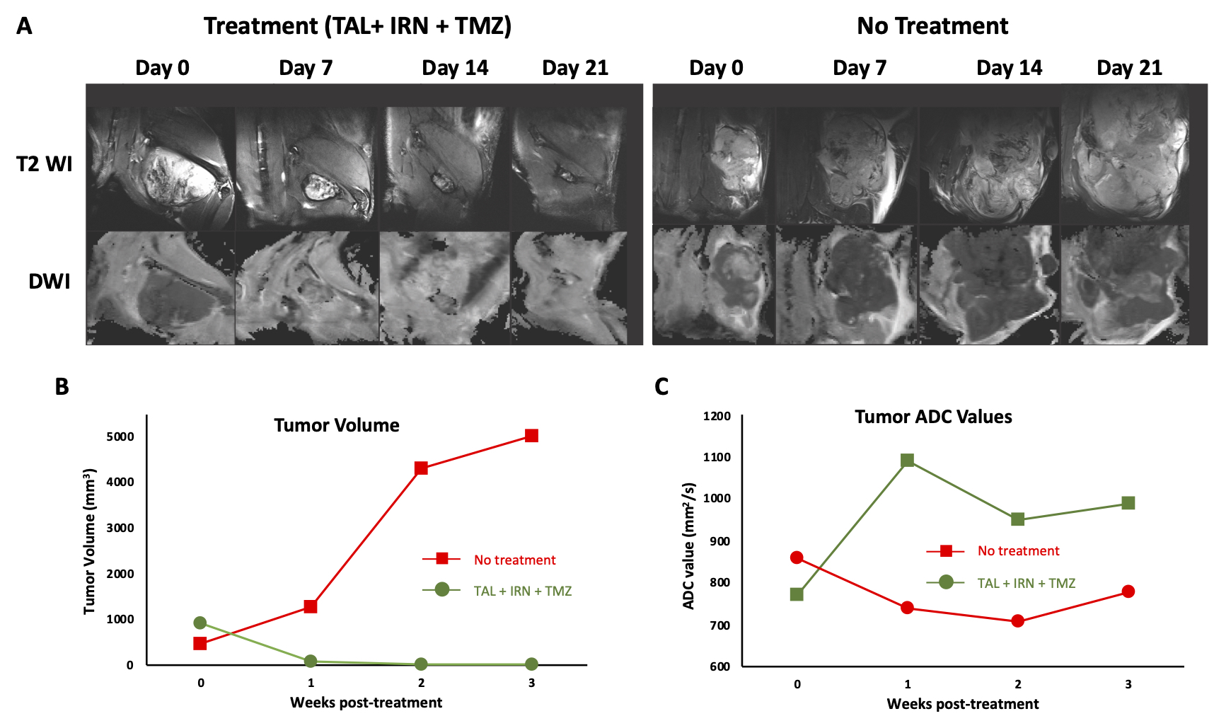
Figure 2. A) T2- and Diffusion-weighted images in the representative TAL + IRN + TMZ treated and untreated mice. Anatomical images indicate the increase in tumor size of the untreated mouse while the tumor is barely visible in the treatment group. B) Measured tumor volume from the multi-slice T2-weighted MRI, and C) measured tumor ROI ADC values in both groups.
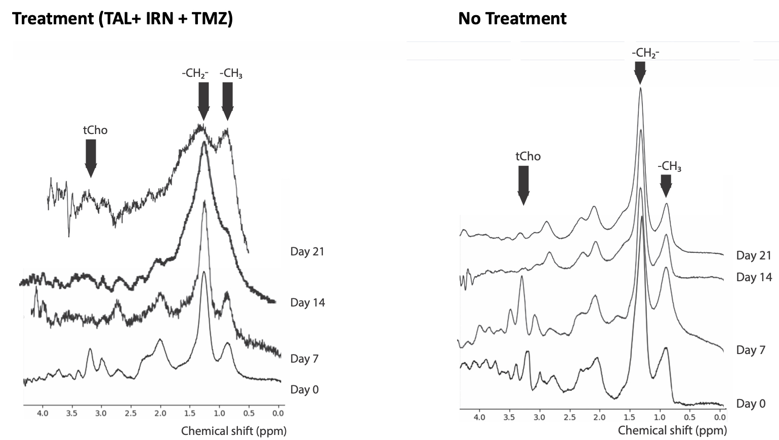
Figure 3. Longitudinal 1H MR spectra from a representative TAL+IRN+TMZ treated mouse and an untreated mouse showing distinct macromolecule peak patterns in the two groups.