Yongsheng Chen1, Jiani Hu2, Yimin Shen2, Lara M Fahmy3,4, Li Zhang3, E. Mark Haacke1,2, and Quan Jiang1,3
1Department of Neurology, Wayne State University School of Medicine, Detroit, MI, United States, 2Department of Radiology, Wayne State University School of Medicine, Detroit, MI, United States, 3Department of Neurology, Henry Ford Health System, Detroit, MI, United States, 4Department of Psychiatry and Behavioral Neurosciences, Wayne State University School of Medicine, Detroit, MI, United States
1Department of Neurology, Wayne State University School of Medicine, Detroit, MI, United States, 2Department of Radiology, Wayne State University School of Medicine, Detroit, MI, United States, 3Department of Neurology, Henry Ford Health System, Detroit, MI, United States, 4Department of Psychiatry and Behavioral Neurosciences, Wayne State University School of Medicine, Detroit, MI, United States
We observed substantial
parenchymal venous participation in cerebral waste clearance in addition to
established cerebrospinal fluid participation.
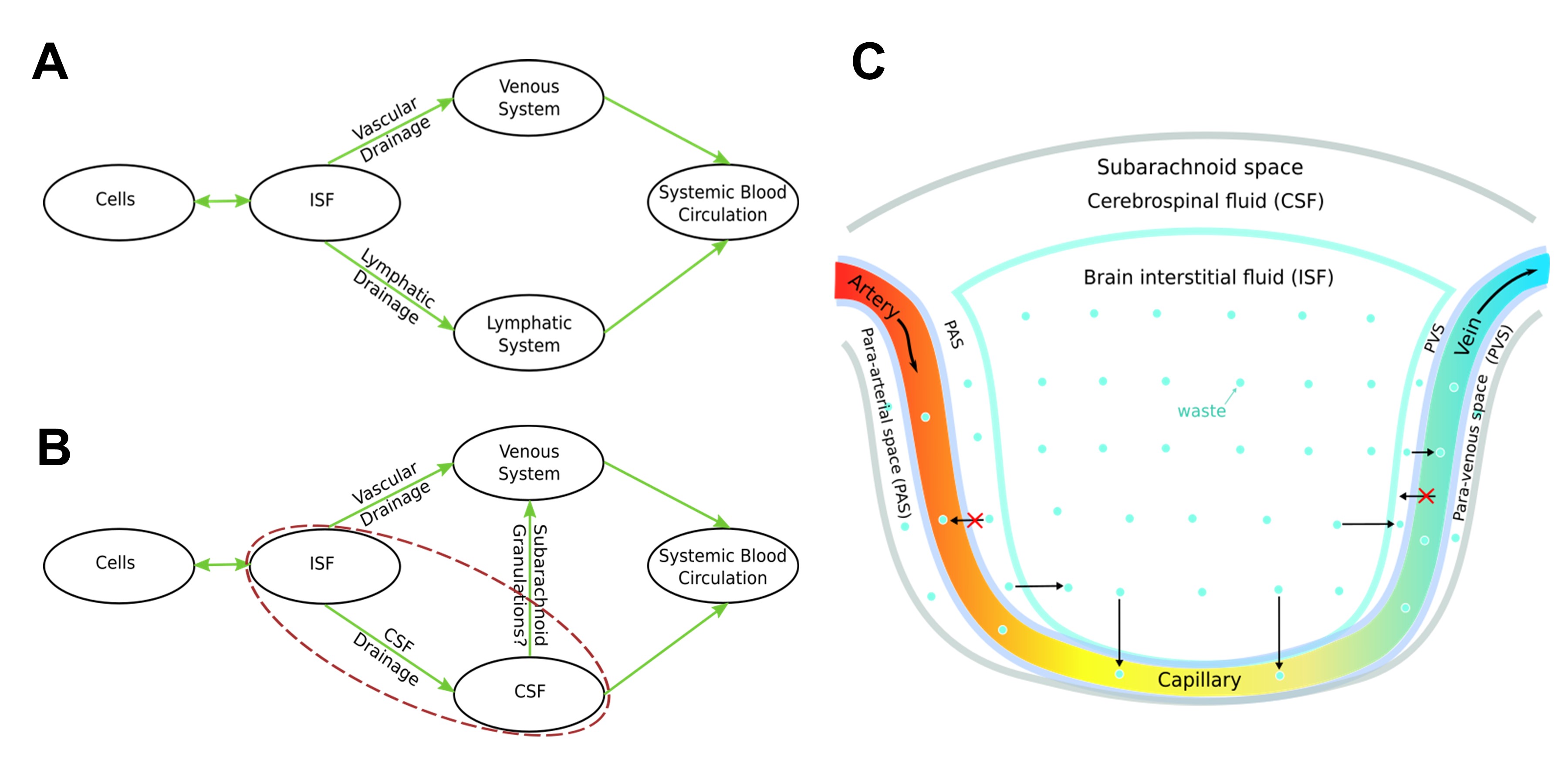
Figure 5. Schemes of waste
clearance. A: Schematic of waste clearance in body tissue outside the
brain. B: Schematic of CWC illustrating the participation of both the
venous system and CSF system; The main difference between CWC and body waste
clearance is the extra layer of CSF (red dashed line in B). C:
Illustration of the one-way transfer of cerebral waster from the brain
parenchyma to capillaries or venules as well as much smaller diameter of the
para-venous pathway comparing to the corresponding venous pathway, which makes
it less effective in waste clearance anatomically.
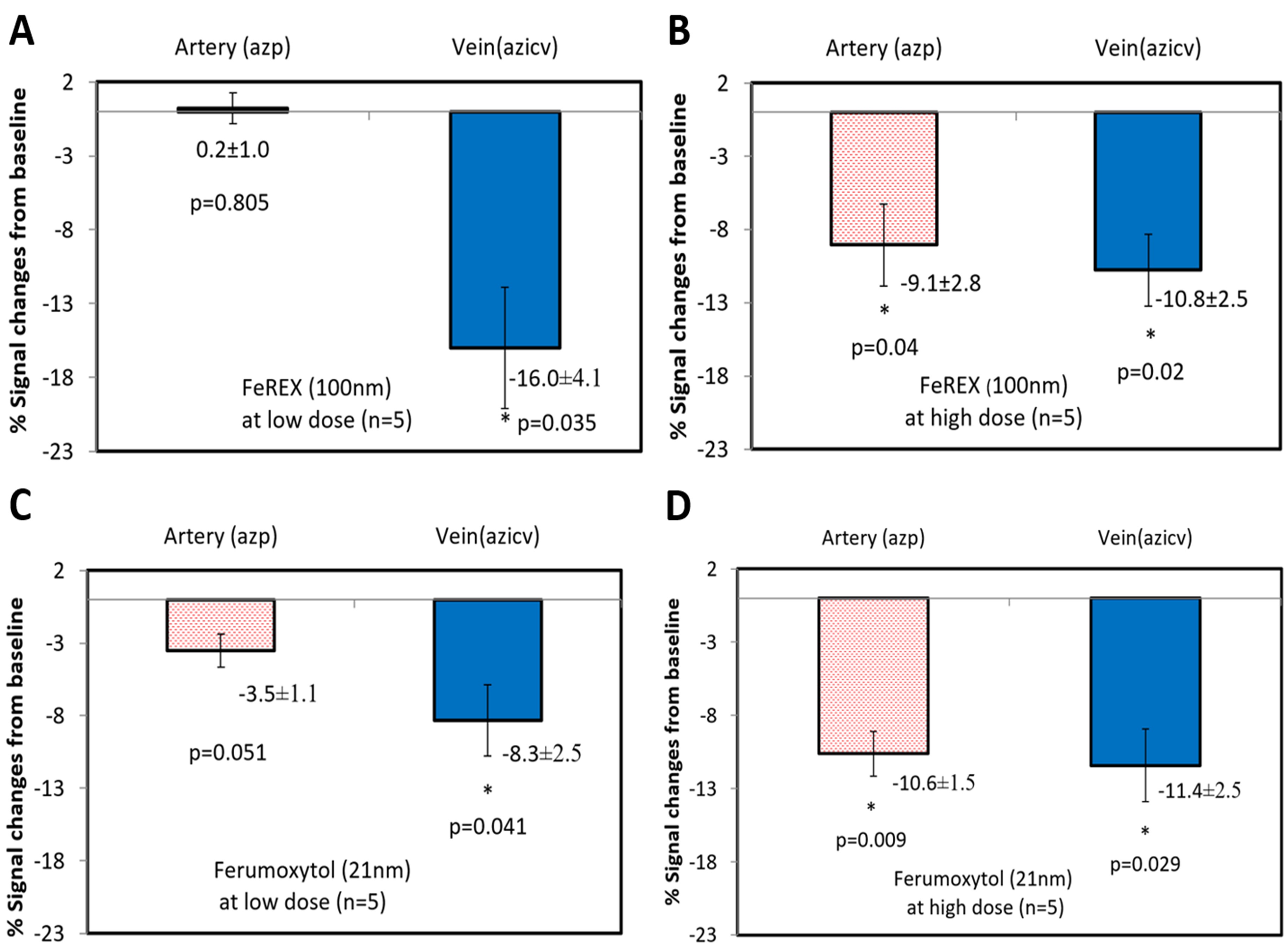
Figure 4.
Participation of the parenchymal venous pathway in CWC. SWI signal intensity
changes post-SPIO CSF tracer infusion in normal rats. A-B: Quantitative
MRI signal intensity changes pre-/post-low dose 75μg (A) and high dose
270 μg (B) FeREX (100nm) in azp and azicv; demonstrating CSF tracer
presence in azicv, but not azp, at low dose. C-D: Quantitative MRI
signal intensity changes pre-/post-low dose 75μg (C) and high dose 240
μg (D) Ferumoxytol (21nm) in azp and azicv; demonstrating CSF tracer
presence in azicv, but not azp, at low dose.