Alexander Saunders1,2, Stefan Bluml1,2, Kevin S King3, and Matthew Borzage2,4
1Radiology, Children's Hospital Los Angeles, Los Angeles, CA, United States, 2Rudi Schulte Research Insitute, Santa Barbara, CA, United States, 3Barrow Neurological Institute, Phenoix, AZ, United States, 4Children's Hospital Los Angeles, Los Angeles, CA, United States
1Radiology, Children's Hospital Los Angeles, Los Angeles, CA, United States, 2Rudi Schulte Research Insitute, Santa Barbara, CA, United States, 3Barrow Neurological Institute, Phenoix, AZ, United States, 4Children's Hospital Los Angeles, Los Angeles, CA, United States
Automatic callosal angle measurement can rapidly and objectively detect patients with normal pressure hydrocephalus. Automatic angle measurement calculated for 4,980 MRIs found that 1.8-4.3% of patients likely had normal pressure hydrocephalus.
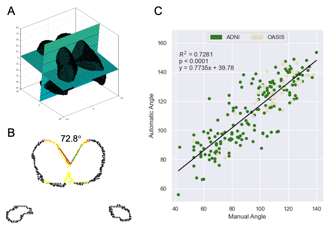
Figure 1 (A) Extracted ventricles with automatically placed oblique axial and coronal reference planes; (B) using the oblique slice from (A), the ventricle walls (black) are sliced, candidate points for the callosal wall are identified (yellow). Refiled points (red) indicate those included in the final calculation. Lines are then fit through the points on each side of the vertex and the angle is calculated; (C) reports the correlation between manual and automated measurements.
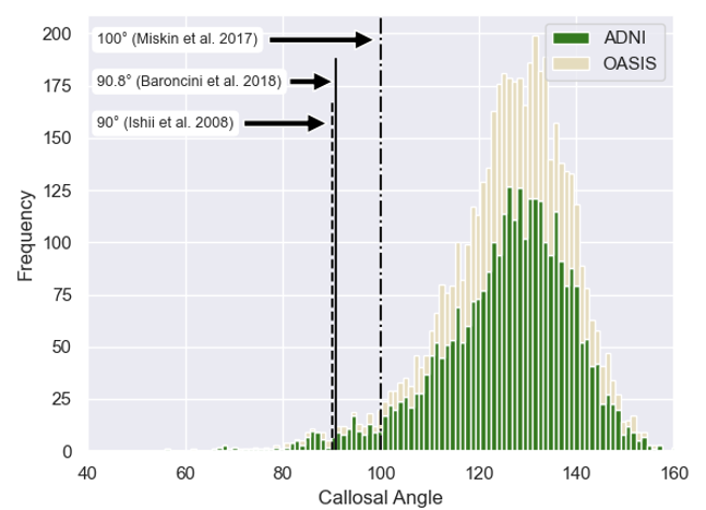
Figure 2 Histogram of 4,980 automatic callosal angle measurements. The data is skewed towards lower angles. Thresholds for suspected normal pressure hydrocephalus are indicated for several publications.