Corinne A. Donnay1, Pauline Maillard1, Charles DeCarli1, and Audrey P. Fan1,2
1Neurology, University of California Davis, Davis, CA, United States, 2Biomedical Engineering, University of California Davis, Davis, CA, United States
1Neurology, University of California Davis, Davis, CA, United States, 2Biomedical Engineering, University of California Davis, Davis, CA, United States
White matter hyperintensities and their surrounding tissue have lower perfusion and higher extracellular water, as reflected by free water, than healthy white matter. Cerebral blood flow increases from lower layers to higher layers. The opposite associations were found with free water.

Figure 2. Summary density plots of all participants (n=19) and clusters (n=331) showing distribution differences between layers 1-4 and WMHs (top) and cortical tissue (bottom). (A) Distribution of CBF values (B) Distribution of FW values
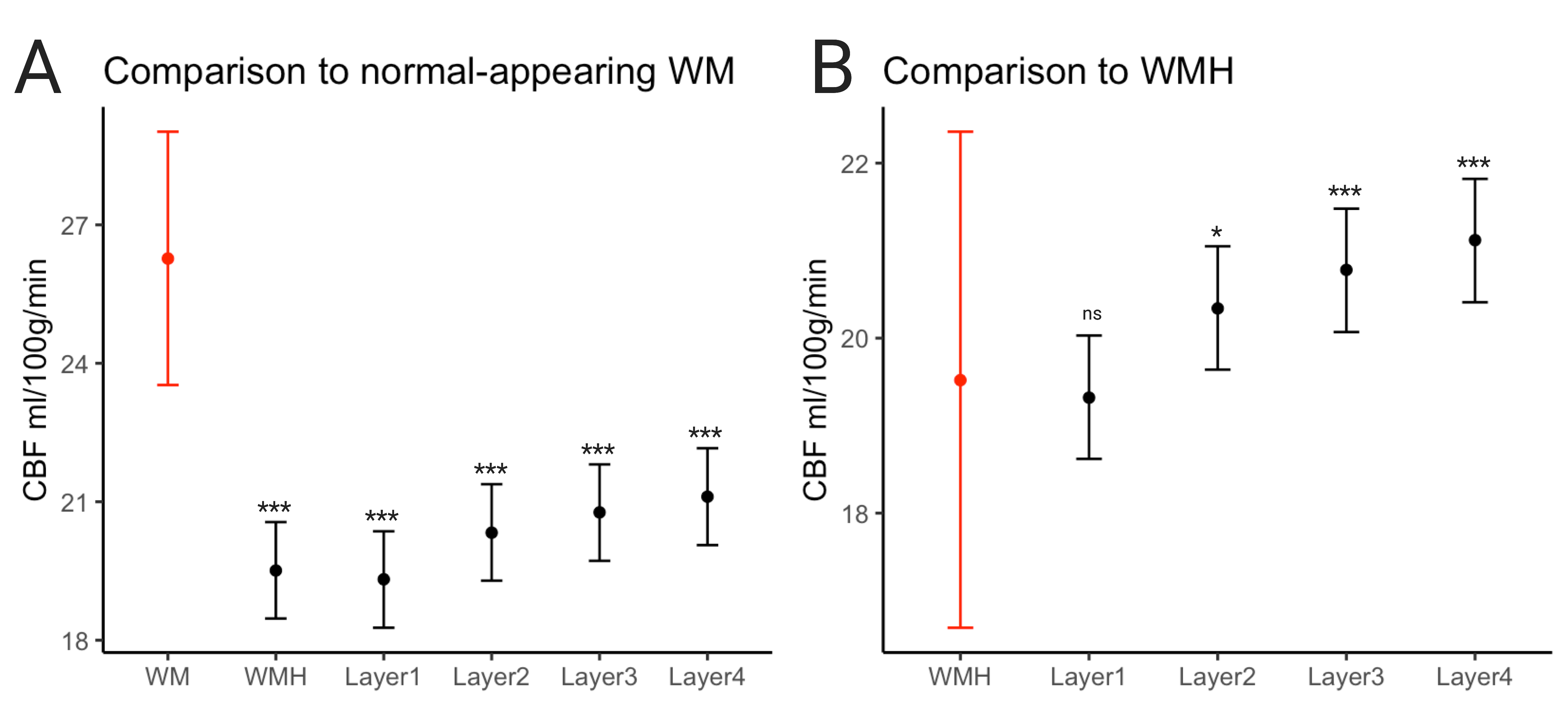
Figure 4. Results of linear mixed model comparing the mean CBF values of (A) WMHs and layers 1-4 to normal-appearing white matter, indicated in red and (B) layers 1-4 to WMHs, indicated in red.
***p<0.001, *p<0.01