Chenyang Li1,2, Marco Muccio1, Dengrong Jiang3, Peiying Liu3, Jiangyang Zhang1, Arjun Masurkar4, Thomas Wisniewski4, Hanzhang Lu3, and Yulin Ge1
1Department of Radiology, Center for Biomedical Imaging, NYU Grossman School of Medicine, New York, NY, United States, 2Vilcek Institute of Graduate Biomedical Sciences, NYU Grossman School of Medicine, New York, NY, United States, 3Department of Radiology and Radiological Science, Johns Hopkins University, Baltimore, MD, United States, 4Department of Neurology, NYU Grossman School of Medicine, New York City, NY, United States
1Department of Radiology, Center for Biomedical Imaging, NYU Grossman School of Medicine, New York, NY, United States, 2Vilcek Institute of Graduate Biomedical Sciences, NYU Grossman School of Medicine, New York, NY, United States, 3Department of Radiology and Radiological Science, Johns Hopkins University, Baltimore, MD, United States, 4Department of Neurology, NYU Grossman School of Medicine, New York City, NY, United States
We implemented advanced neurovascular MRI techniques to evaluate the white matter hemodynamics including cerebral blood flow (CBF) and cerebrovascular reactivity (CVR) in patients with varying degrees of WMHs. Lower CBF and CVR function could be potentially associated with WMHs.
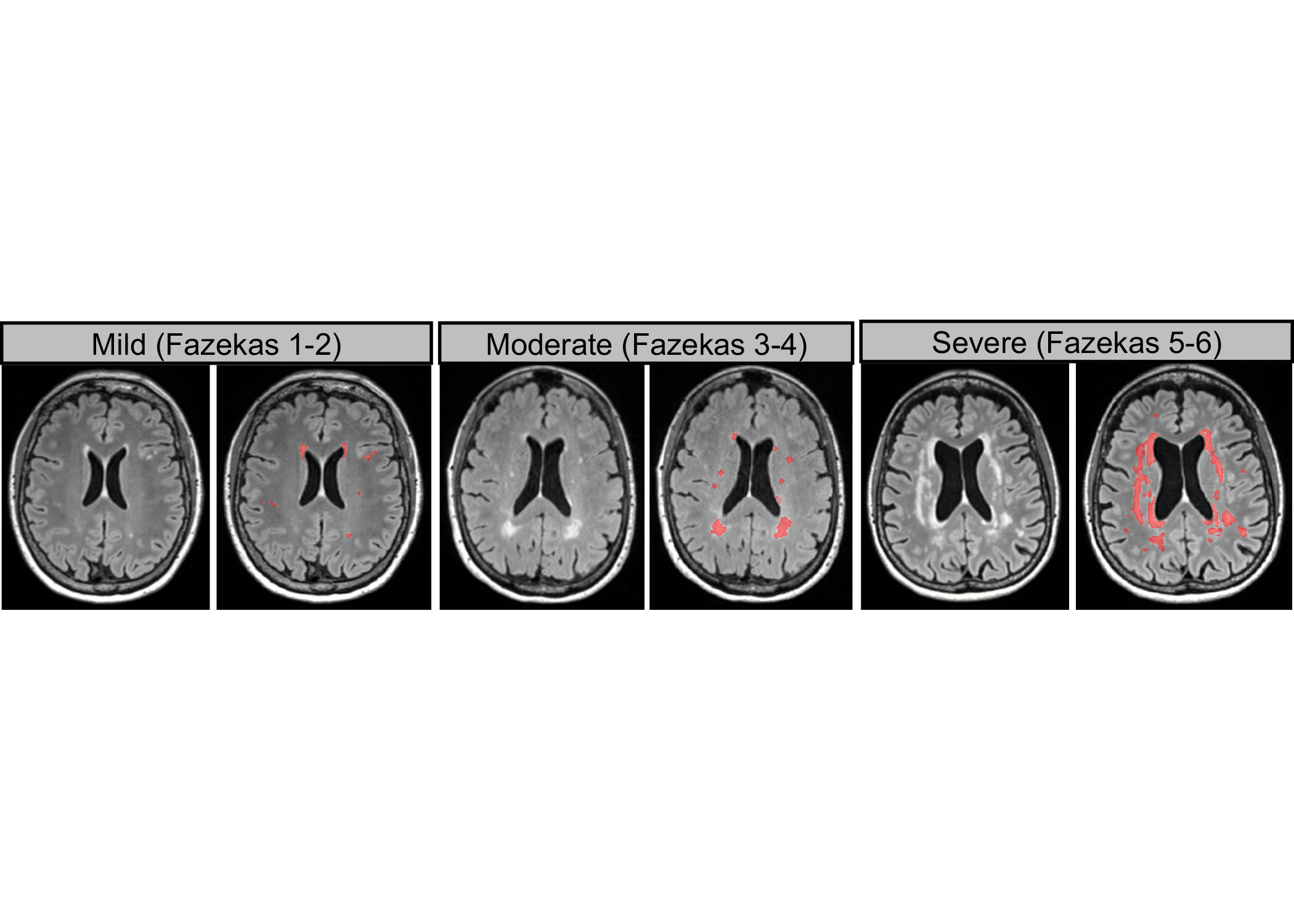
Figure 1.
Representation WMHs lesions on T2-FLAIR images in mild,
moderate and severe lesion groups and its corresponding lesion segmentation result (delineated in red).
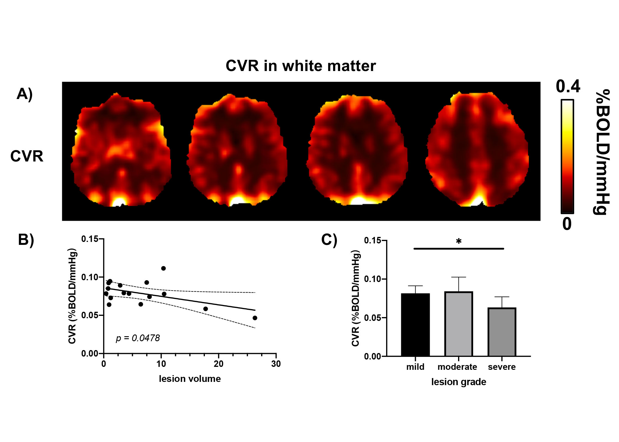
Figure 2.
A)
Representation of CVR maps in WMHs patients. B) Correlation analysis between lesion volume and white matter CVR. C) Group-wise comparison of white matter CVR between mild, moderate and severe lesion groups.