Miller Fawaz1, Sara Gharabaghi1, Mojtaba Jokar1, Ying Wang1,2, Chao Chai3, and E. Mark Haacke1,2
1Magnetic Resonance Innovations, Inc., Bingham Farms, MI, United States, 2Wayne State University, Detroit, MI, United States, 3Tianjin First Central Hospital, Tianjin, China
1Magnetic Resonance Innovations, Inc., Bingham Farms, MI, United States, 2Wayne State University, Detroit, MI, United States, 3Tianjin First Central Hospital, Tianjin, China
We improved our existing pipeline for automatic
cerebral microbleed detection by adding a false positive correction step using STAGE
imaging. The
sensitivity reached 92.3 with 3.7 false positives per case on average, creating
a clinically viable STAGE imaging based microbleed detection.
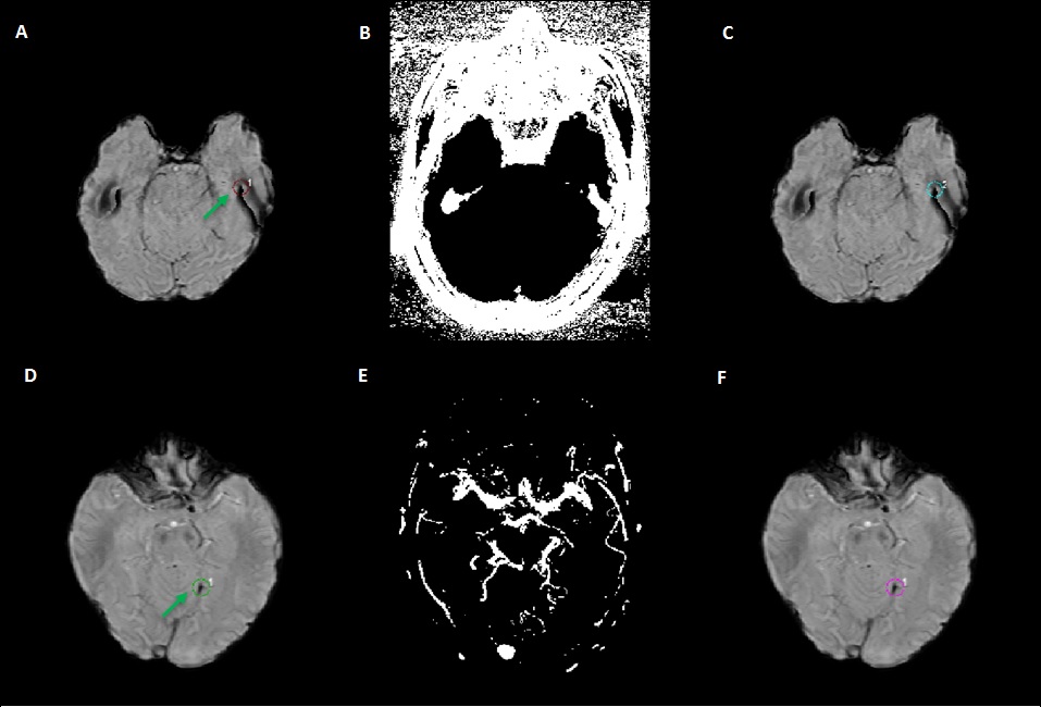
Figure 1. Two
Stroke STAGE cases with corrected FPs. First case (1st row) has a
detected CMB (shown in red circle in A) on the SWI (A) that was located on the
edge, and later using the extracted edge mask (B), it was removed from the SWI
(C) as a false positive shown with cyan circle. Second case (2nd
row) shows a detected CMB (shown in green circle) on the SWI (D) that was
located on the vein, and later using the extracted vein mask (E), it was
removed from the SWI (F) as a false positive shown with purple circle. Those
are false positives that would have otherwise been included in the result.
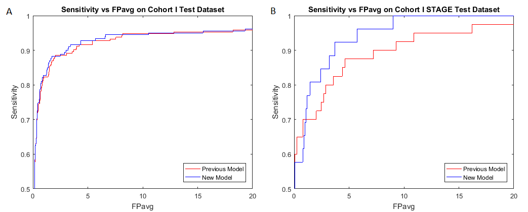
Figure 2.
Comparison of new and old pipelines on all datasets (STAGE and non-STAGE) (A)
and only STAGE datasets (B) of the previously tested cohort. As seen in the
figure, the new model works better on both datasets, and there is a performance
improvement on STAGE datasets.