Kathleen B Miller1, Leonardo A Rivera-Rivera1, Oliver Wieben1, Kevin M Johnson1, Sterling C Johnson1, and Jill N Barnes1
1University of Wisconsin-Madison, Madison, WI, United States
1University of Wisconsin-Madison, Madison, WI, United States
Low frequency oscillations (LFOs) of intracranial vessels were measured using real time 4D flow MRI at baseline and during hypercapnia. Hypercapnia increased LFOs in the superior sagittal sinus with no changes observed in the internal carotid arteries.
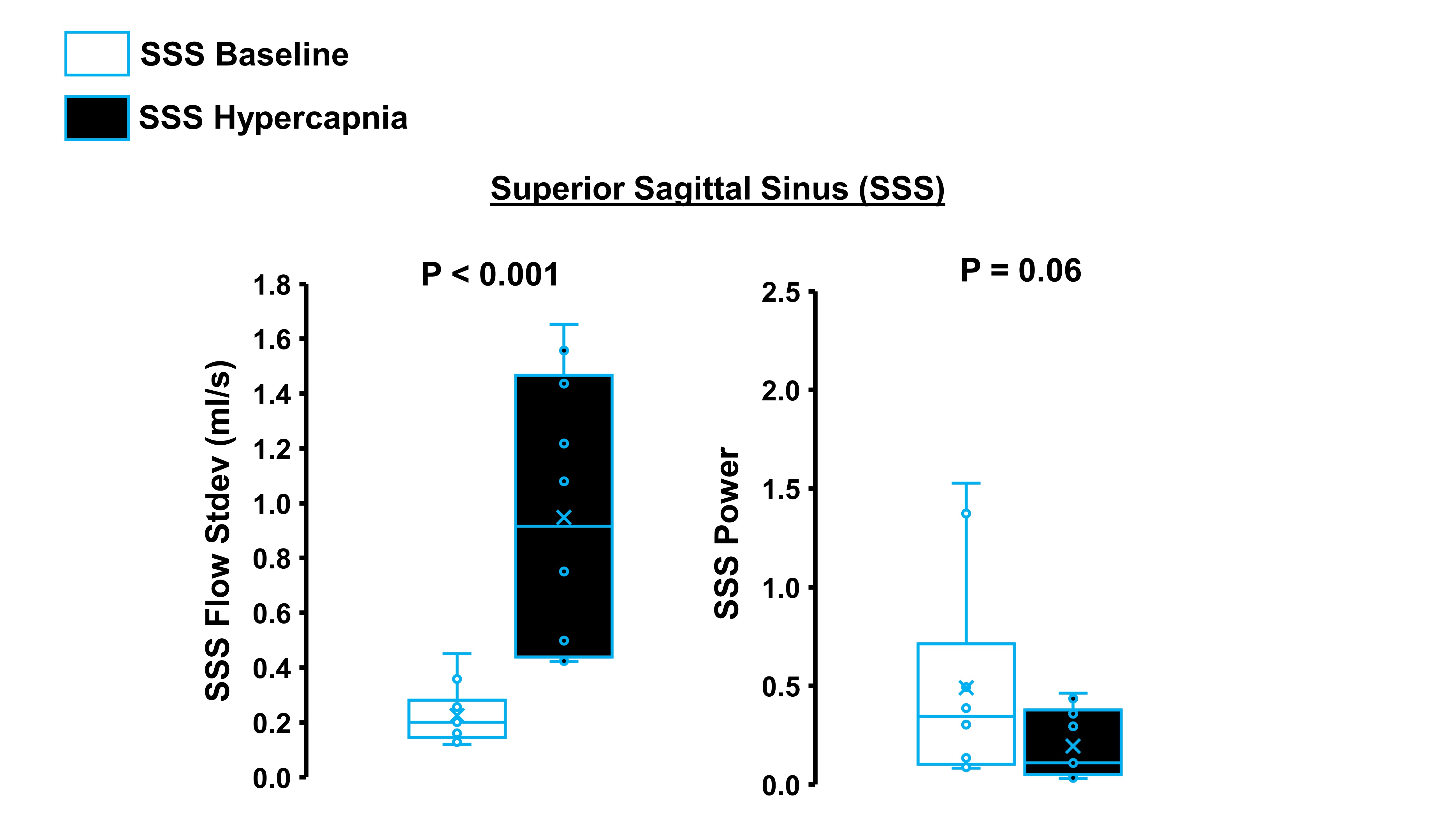
Figure 3 shows box plots of the flow standard deviation (stdev) and the average low-frequency power measured in the superior sagittal sinus (SSS) (n=10). The baseline condition is shown in white and the hypercapnic condition is shown in black. Hypercapnia was associated with a significant increase in flow standard deviation. There was also a trend for lower, low-frequency power in the hypercapnic condition compared with baseline.
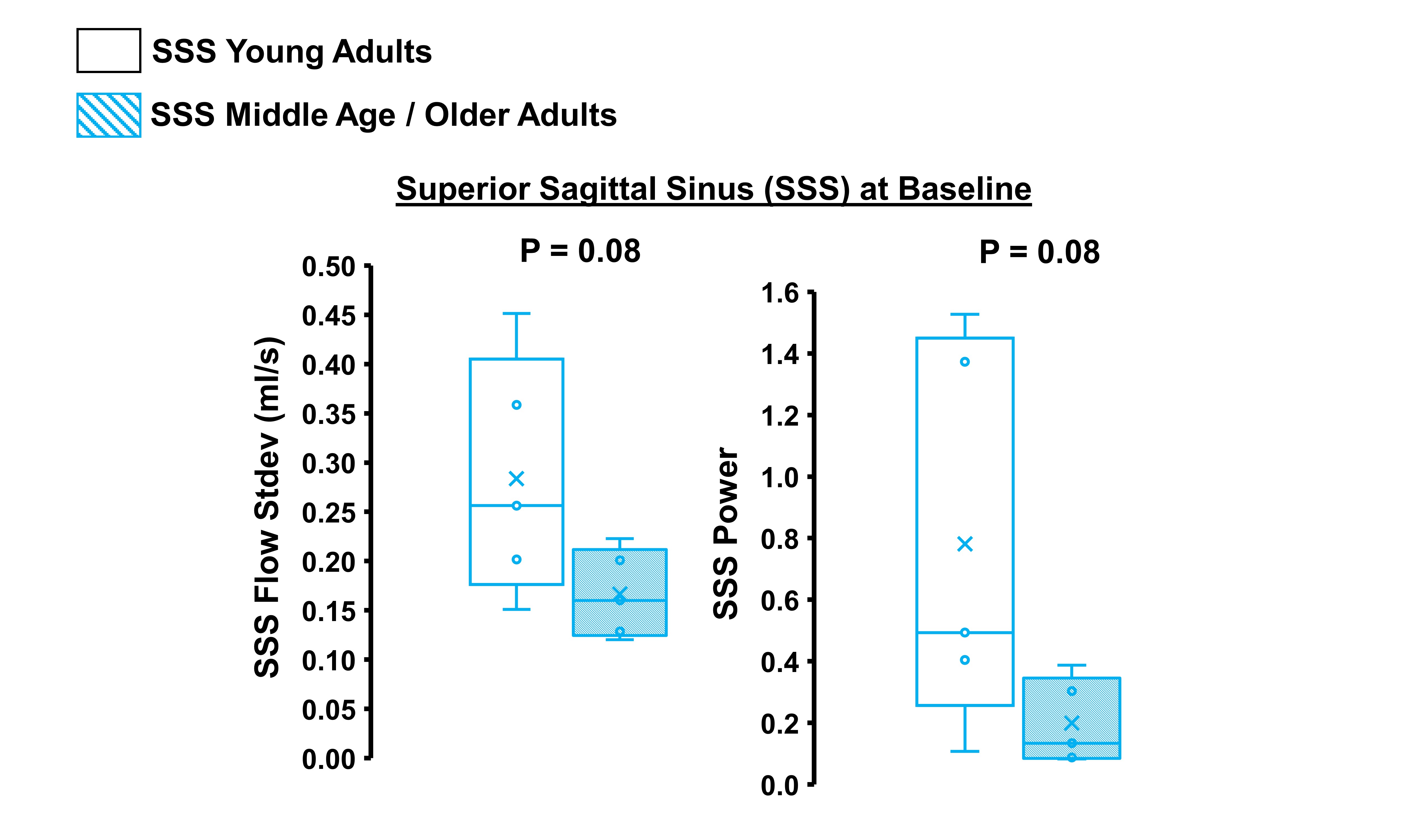
Figure 4 shows box plots of the flow standard deviation (stdev) and the average low-frequency power measured in the superior sagittal sinus (SSS) at baseline. Young adults (n=5) are shown in white and middle-age/older adults (n=5) are shown in dashed blue lines. There was a trend for middle-age/older adults to demonstrate a smaller flow standard deviation, and lower, low-frequency power compared with young adults.