Satoshi Yatsushiro1, Tomohiko Horie2, Mitsunori Matsumae3, and Kagayaki Kuroda1
1Department of Human and Information Science, Tokai University, Hiratsuka, Japan, 2Department of Radiological Technology, Tokai University Hospital, Isehara, Japan, 3Department of Neurosurgery, Tokai University, Isehara, Japan
1Department of Human and Information Science, Tokai University, Hiratsuka, Japan, 2Department of Radiological Technology, Tokai University Hospital, Isehara, Japan, 3Department of Neurosurgery, Tokai University, Isehara, Japan
Asynchronous phase contrast with Stockwell transform and power mapping characterized the spatial difference between the cardiac- and respiratory-driven CSF motion in the intracranial space under free breathing.
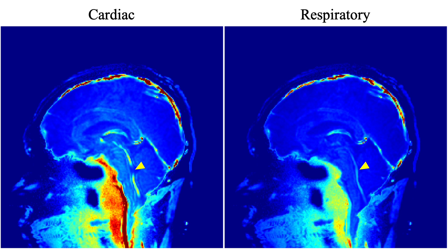
Figure 3. Power maps of the cardiac- and respiratory-driven CSF motion components. The power of the cardiac-driven component looked like higher around the brainstem than that of the respiratory. Meanwhile, in the fourth ventricle, the contribution of the respiratory-driven component was high indicated by yellow arrow head.
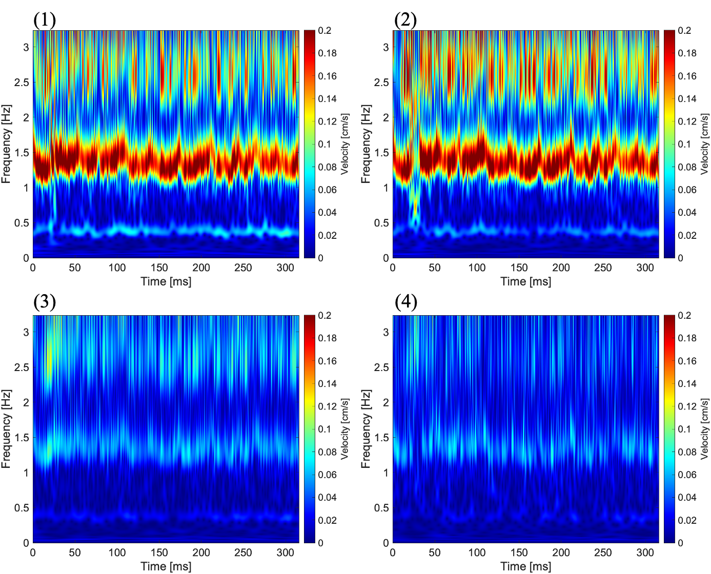
Figure 2. Spectrograms of CSF velocity obtained from ROIs shown in Figure 1. Number at top-left on each image corresponds to the ROI number in Figure 1. Cardiac- and respiratory-driven velocities gradually changes from (1) to (4).