Khin Khin Tha1,2, Yuta Urushibata3, Hiroyuki Hamaguchi2, and Hideki Hyodoh4
1Global Center for Biomedical Science and Engineering, Hokkaido University Faculty School of Medicine, Sapporo, Japan, 2Department of Biomarker Imaging Science, Hokkaido University Graduate School of Biomedical Science and Engineering, Sapporo, Japan, 3Siemens Healthcare K.K., Tokyo, Japan, 4Department of Forensic Medicine, Hokkaido University Faculty of Medicine, Sapporo, Japan
1Global Center for Biomedical Science and Engineering, Hokkaido University Faculty School of Medicine, Sapporo, Japan, 2Department of Biomarker Imaging Science, Hokkaido University Graduate School of Biomedical Science and Engineering, Sapporo, Japan, 3Siemens Healthcare K.K., Tokyo, Japan, 4Department of Forensic Medicine, Hokkaido University Faculty of Medicine, Sapporo, Japan
In this ex vivo
CSF analysis by CEST MRI, the normalized area for intermediate exchanging amines showed a weak positive correlation with the CSF protein
concentration and specific gravity and a weak negative correlation with pH.
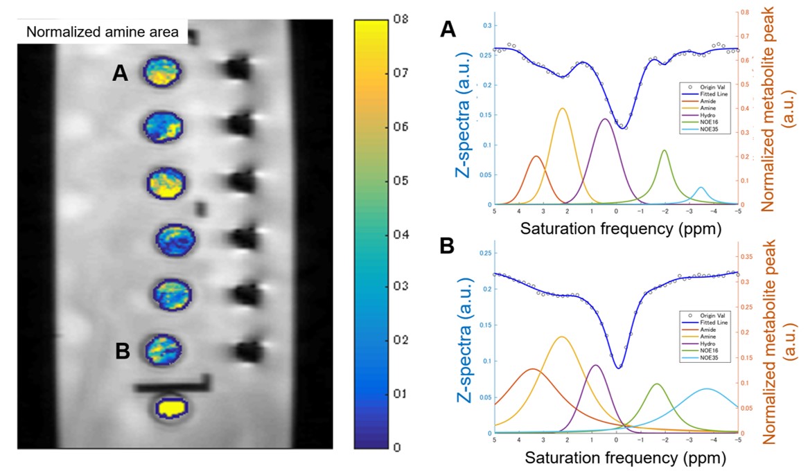
Fig 5. Maps of
the normalized area of intermediate exchanging amines for six CSF and one control phantoms (left). Note the variation between two labeled test tubes -- test tube "A" (CSF protein concentration= 487 mg/dL) and test tube "B" (177 mg/dL). "A" has a higher normalized area than "B". The corresponding z spectra (right) are
also given.
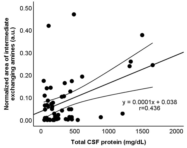
FIg 2. Scatterplots showing the correlation between the
normalized area for intermediate exchanging amines and CSF protein
concentration (r=0.436, P=0.001). The straight and curved lines indicate the mean
and 95% confidence interval.