Dennis Thomas1,2, Ana-Maria Oros-Peusquens1, Dirk Poot3, and N. Jon Shah1,4,5,6
1Institute of Neuroscience and Medicine-4, Forschungszentrum Jülich, Jülich, Germany, 2RWTH Aachen University, Aachen, Germany, 3Department of Radiology and Nuclear medicine, Erasmus Medical Center, Rotterdam, Netherlands, 4Department of Neurology, RWTH Aachen University, Aachen, Germany, 5Institute of Neuroscience and Medicine 11, INM-11, JARA, Forschungszentrum Jülich, Jülich, Germany, 6JARA - BRAIN - Translational Medicine, Aachen, Germany
1Institute of Neuroscience and Medicine-4, Forschungszentrum Jülich, Jülich, Germany, 2RWTH Aachen University, Aachen, Germany, 3Department of Radiology and Nuclear medicine, Erasmus Medical Center, Rotterdam, Netherlands, 4Department of Neurology, RWTH Aachen University, Aachen, Germany, 5Institute of Neuroscience and Medicine 11, INM-11, JARA, Forschungszentrum Jülich, Jülich, Germany, 6JARA - BRAIN - Translational Medicine, Aachen, Germany
A method to obtain high-resolution, isotropic, whole-brain water content maps within a clinically-relevant acquisition time has been developed and evaluated on a carrageenan phantom. In vivo, whole-brain results from a volunteer are presented.
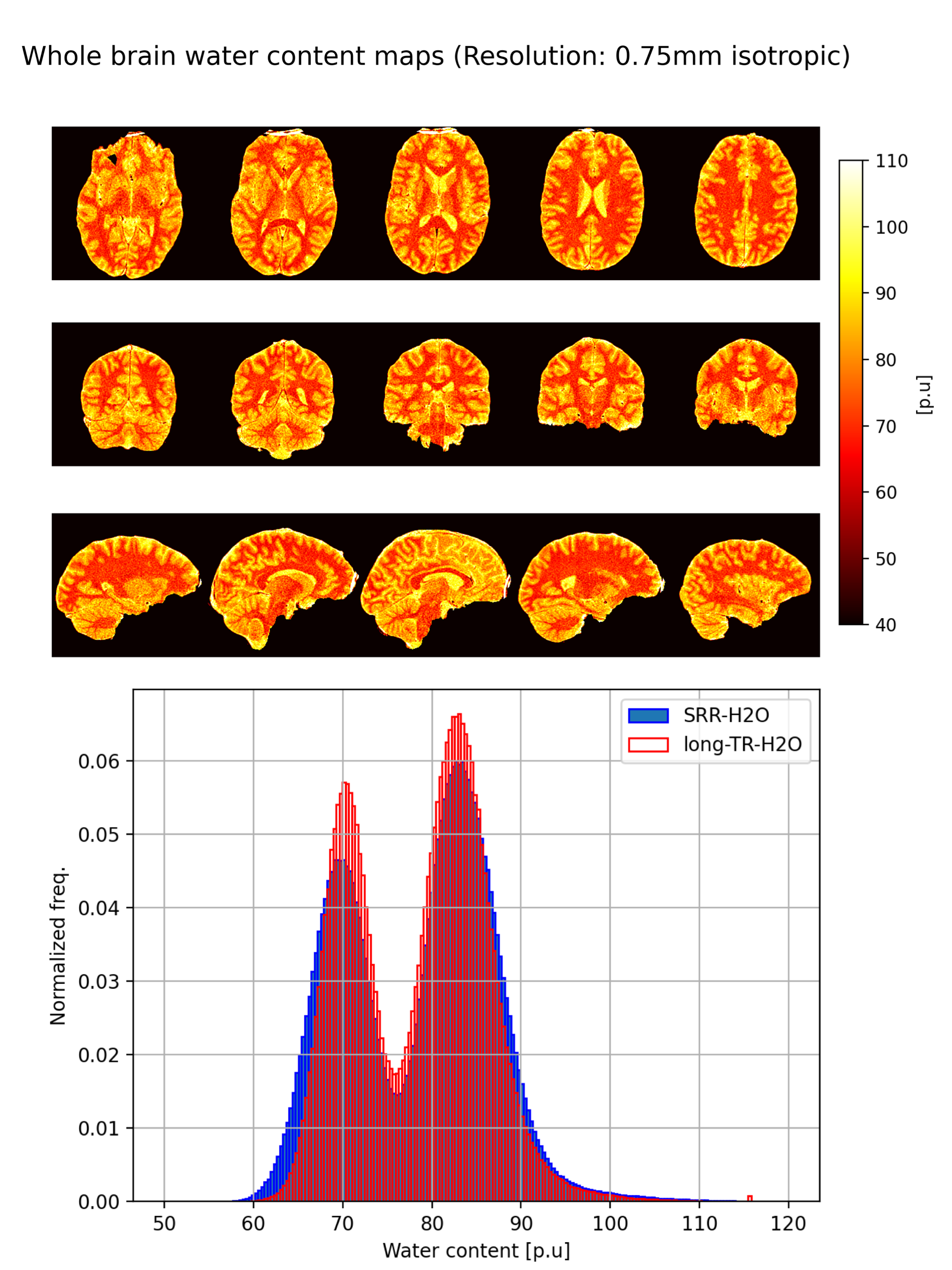
Figure 4: Upper figure: Different slices of the whole brain H2O maps (in percentage units) generated with the proposed method are displayed in the transverse, coronal and sagittal planes (from top to bottom). The water content is calibrated to that of the CSF which is assumed to have 100% water content. A CSF T1 value of 4300ms was used for calibration. Lower figure: Histogram plots of the SRR-H2O and long-TR-H2O values. The peak at around 70% is for the WM and that around 83% is for the GM.
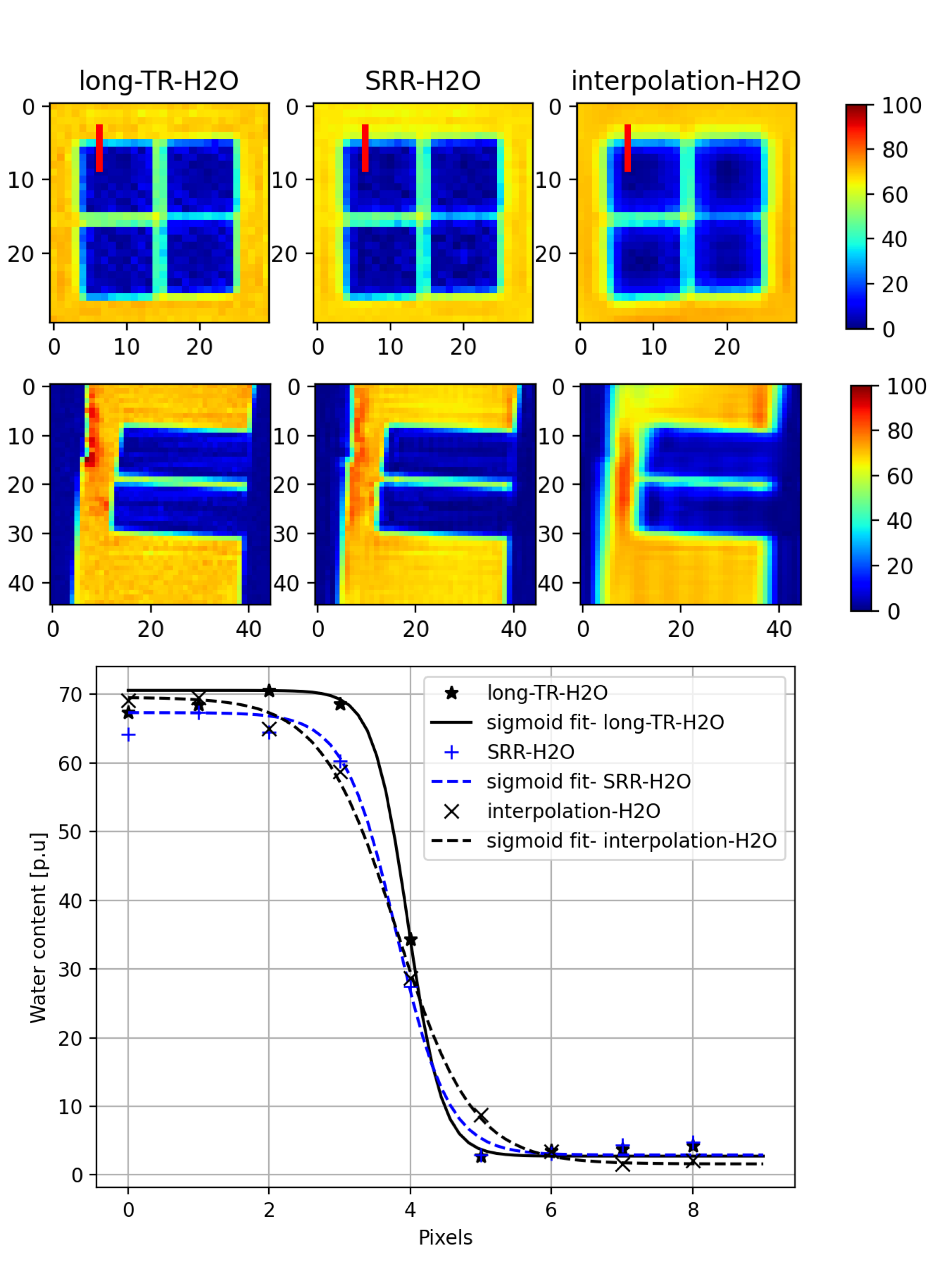
Figure 3: Upper rows show water content maps (in percentage units) of an ROI in the phantom obtained with reference (long-TR-H2O), proposed (SRR-H2O) and the zero padded interpolation (Interpolation-H2O) methods obtained with an equal scan time. Blurring can be noticed in the interpolation-H2O method while the SRR-H2O maps are comparable to the reference method. The lower figure plots the sigmoid fit to an edge selected which is shown as a red line in the top most figure. The steeper the fitted curve, the better the resolution.