Adam C Richie-Halford1, Jason Yeatman2, Noah Simon3, and Ariel Rokem4
1eScienceInstitute, University of Washington, Seattle, WA, United States, 2Graduate School of Education and Division of Developmental and Behavioral Pediatrics, Stanford University, Stanford, CA, United States, 3Department of Biostatistics, University of Washington, Seattle, WA, United States, 4Department of Psychology, University of Washington, Seattle, WA, United States
1eScienceInstitute, University of Washington, Seattle, WA, United States, 2Graduate School of Education and Division of Developmental and Behavioral Pediatrics, Stanford University, Stanford, CA, United States, 3Department of Biostatistics, University of Washington, Seattle, WA, United States, 4Department of Psychology, University of Washington, Seattle, WA, United States
We introduce a novel method for analysis of diffusion MRI (dMRI) tractometry data, based on grouped penalized regression, that provides both accurate prediction of phenotypic information and results that are readily interpretable.
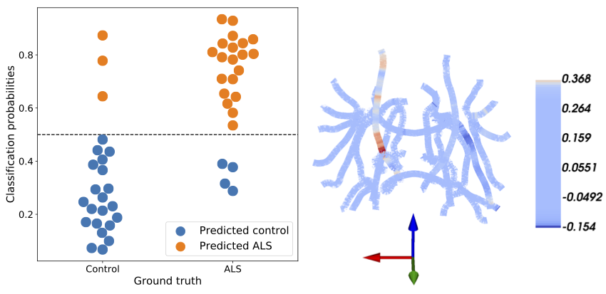
Figure 2: SGL accurately predicts ALS. Left: classification probabilities for each
subject’s ALS diagnosis. Controls are on the left, patients are on the right, predicted controls are in blue, and predicted patients are in orange. The SGL algorithm achieves 86% accuracy. Right: SGL coefficients are presented on a skeleton of the major white matter fiber tracts. The
brain is oriented with the right hemisphere to our left and anterior out of the page. As
expected large coefficients are in the fractional anisotropy of the corticospinal tract.
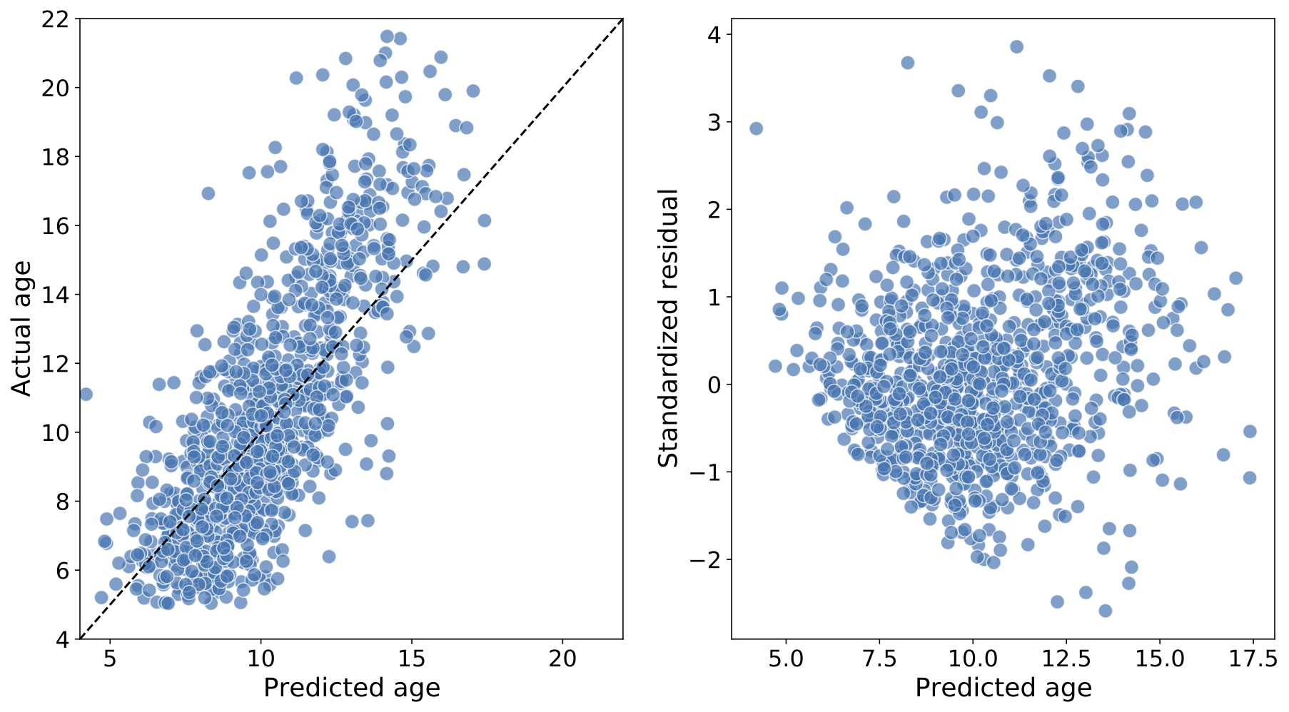
Figure 4: Predicting age in the Healthy Brain Network, a large pediatric neuroimaging study. Left: The predicted age of each individual (on the abscissa) and true age (on the ordinate), from the test splits (i.e., when each subject’s data was held out in fitting the model); an accurate prediction falls close to the $$$y = x$$$ line (dashed). The mean absolute error in this case is 1.5 years and R2 = 0.56. Right: Standardized residuals (on the abscissa) as a function of the true age (on the ordinate). Predictions are generally more accurate for younger individuals.