Sophie You1, Evan M. Masutani1, Joy Liau2, Marcus T. Alley3, Shreyas S. Vasanawala3, and Albert Hsiao2
1School of Medicine, University of California, San Diego, La Jolla, CA, United States, 2Department of Radiology, University of California, San Diego, La Jolla, CA, United States, 3Department of Radiology, Stanford University School of Medicine, Stanford, CA, United States
1School of Medicine, University of California, San Diego, La Jolla, CA, United States, 2Department of Radiology, University of California, San Diego, La Jolla, CA, United States, 3Department of Radiology, Stanford University School of Medicine, Stanford, CA, United States
Using a multichannel 3D convolutional neural network, we demonstrate the feasibility of automating background phase error correction in abdominopelvic 4D Flow MRI. Quantitative improvements in flow consistency are comparable to manual correction.
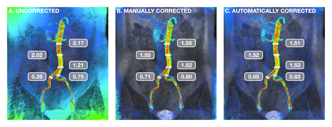
FIGURE 4: Visual example of background phase-error correction. Coronal view of the aorta and common iliac arteries during peak systole, with flow velocity represented by a colormap ranging from red (80 cm/s) to blue (0 cm/s). For assessment of flow continuity, measurements (in L/min) were taken at multiple locations. Corrected velocity measurements demonstrated improved consistency along the length of the infrarenal aorta as well as conservation of mass post-bifurcation.
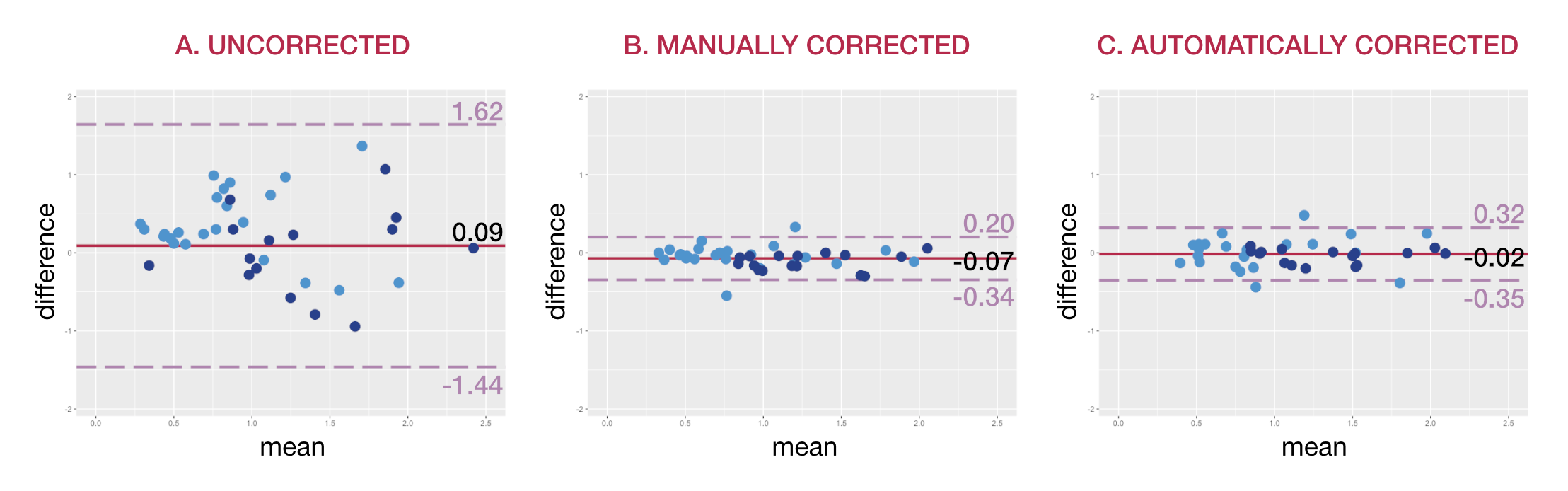
FIGURE 5: Bland-Altman analysis. Consistency of flow measurements are shown for (a) uncorrected velocity data, (b) velocity data corrected manually, and (c) velocity data corrected automatically. Light blue points represent comparisons of arterial and venous flow, while dark blue points represent comparisons of flow before and after vessel bifurcation. Corrected measurements in (b) and (c) demonstrate greater consistency with narrower limits of agreement than seen in (a).