Marina Manso Jimeno1,2, John Thomas Vaughan Jr.1,2, and Sairam Geethanath2
1Columbia University, New York, NY, United States, 2Columbia Magnetic Resonance Research Center (CMRRC), New York, NY, United States
1Columbia University, New York, NY, United States, 2Columbia Magnetic Resonance Research Center (CMRRC), New York, NY, United States
The proposed method for spiral deblurring of fMRI images outperforms MFI at various field inhomogeneity ranges with minimum SSIM of 0.97, pSNR greater than 35 dB, and HFEN smaller than 0.17 and does not require field map calibration.
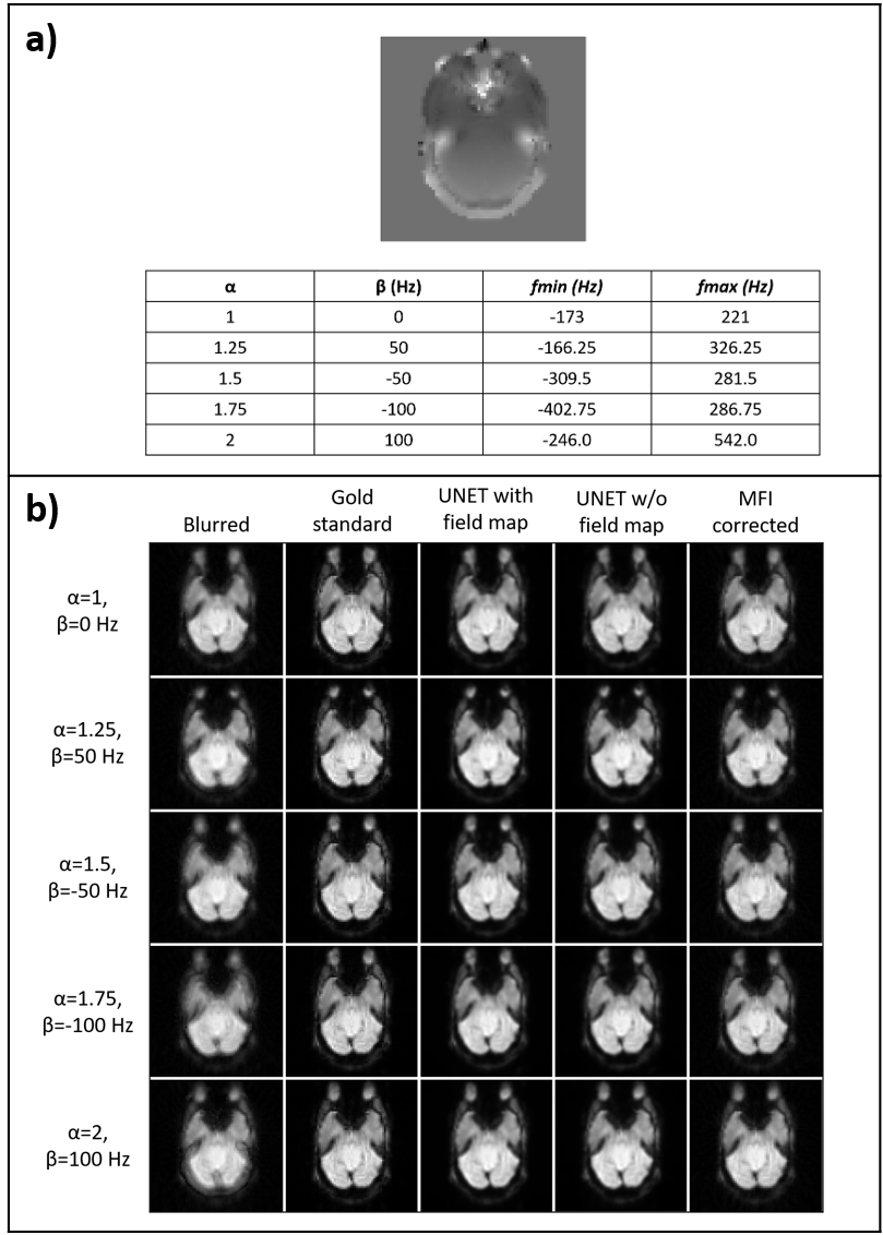
Figure 2. Validation results. Field map augmentation modified the frequency range of each slice depending on the parameters 𝛼 and β. a) shows the different combinations for an example slice and the achieved frequency ranges. b) Image panel displaying the blurred image, gold standard, U-net correction with field map, U-net correction without field map, and MFI correction images.
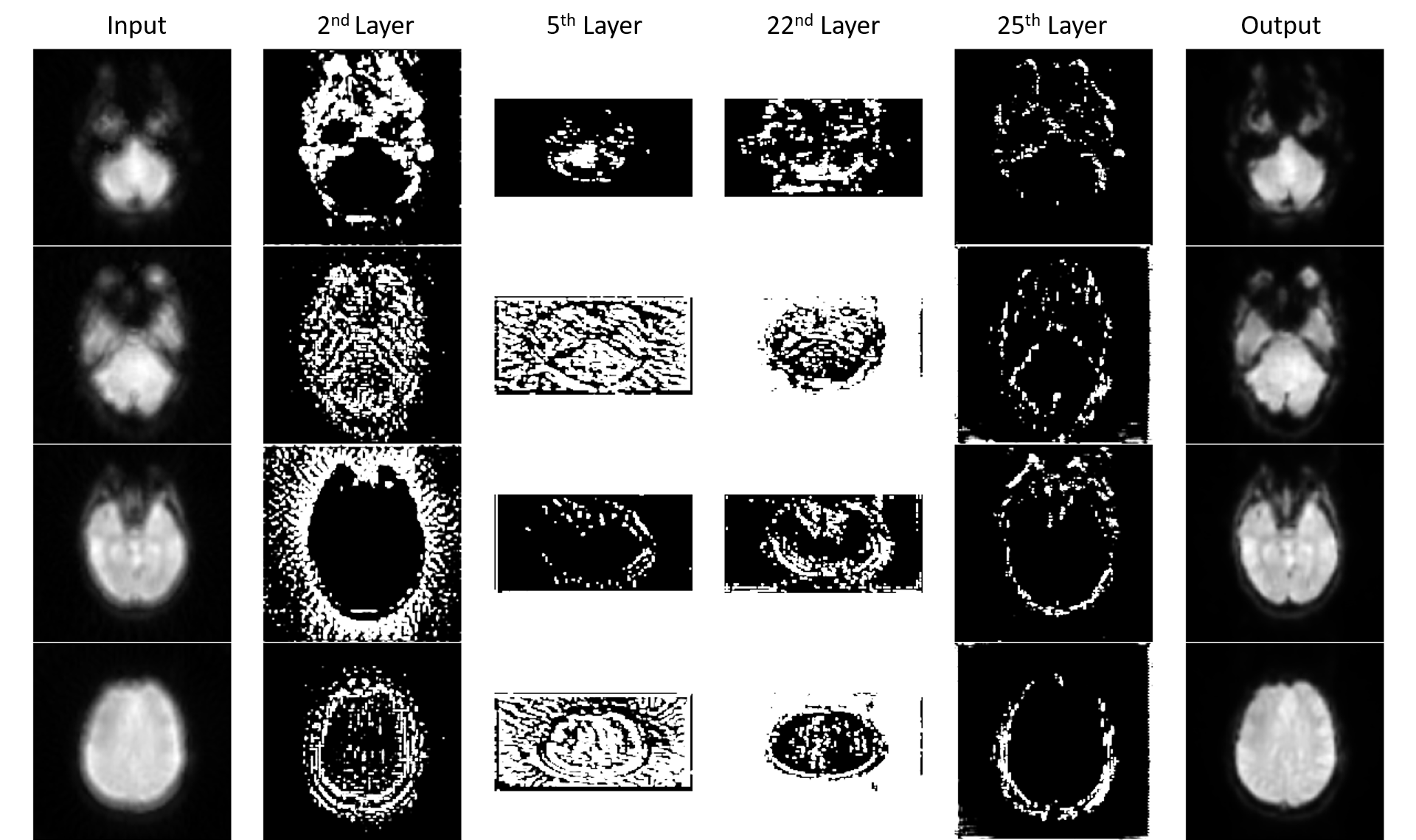
Figure 4. Filter visualization. Visualization of representative filters from the 2nd, 5th, 22nd, and 25th convolutional layers for four different brain slices from the testing dataset for model explainability.