Mario Serrano-Sosa1, Jared Van Snellenberg2, Jiayan Meng2, Jacob Luceno2, Karl Spuhler3, Jodi Weinstein2, Anissa Abi-Dargham2, Mark Slifstein2, and Chuan Huang2,4
1Biomedical Engineering, Stony Brook University, Stony Brook, NY, United States, 2Psychiatry, Renaissance School of Medicine at Stony Brook University, Stony Brook, NY, United States, 3Radiation Oncology, NYU Langone, New York, NY, United States, 4Radiology, Renaissance School of Medicine at Stony Brook University, Stony Brook, NY, United States
1Biomedical Engineering, Stony Brook University, Stony Brook, NY, United States, 2Psychiatry, Renaissance School of Medicine at Stony Brook University, Stony Brook, NY, United States, 3Radiation Oncology, NYU Langone, New York, NY, United States, 4Radiology, Renaissance School of Medicine at Stony Brook University, Stony Brook, NY, United States
Multi-Task Learning provides reliable striatal subregion
segmentations with more comparable PET and fMRI results that closely match those obtained with manually drawn ROIs than atlas-based
segmentations
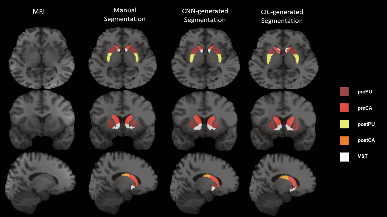
Figure 2.
Transverse (top), coronal (middle) and sagittal (bottom) views of T1-weighted
MR Image, overlaid with ROIs by manual segmentation, MTL-generated
segmentation, and CIC-generated segmentation. Transverse slices of manual
segmentation show postPU to have slightly irregular shape compared to a
smoother postPU in MTL-generated segmentation. Coronal and sagittal slices of
the MRI and manual segmentation also show VST to have irregular shape compared
to a smoothed and more circular VST in MTL-generated.
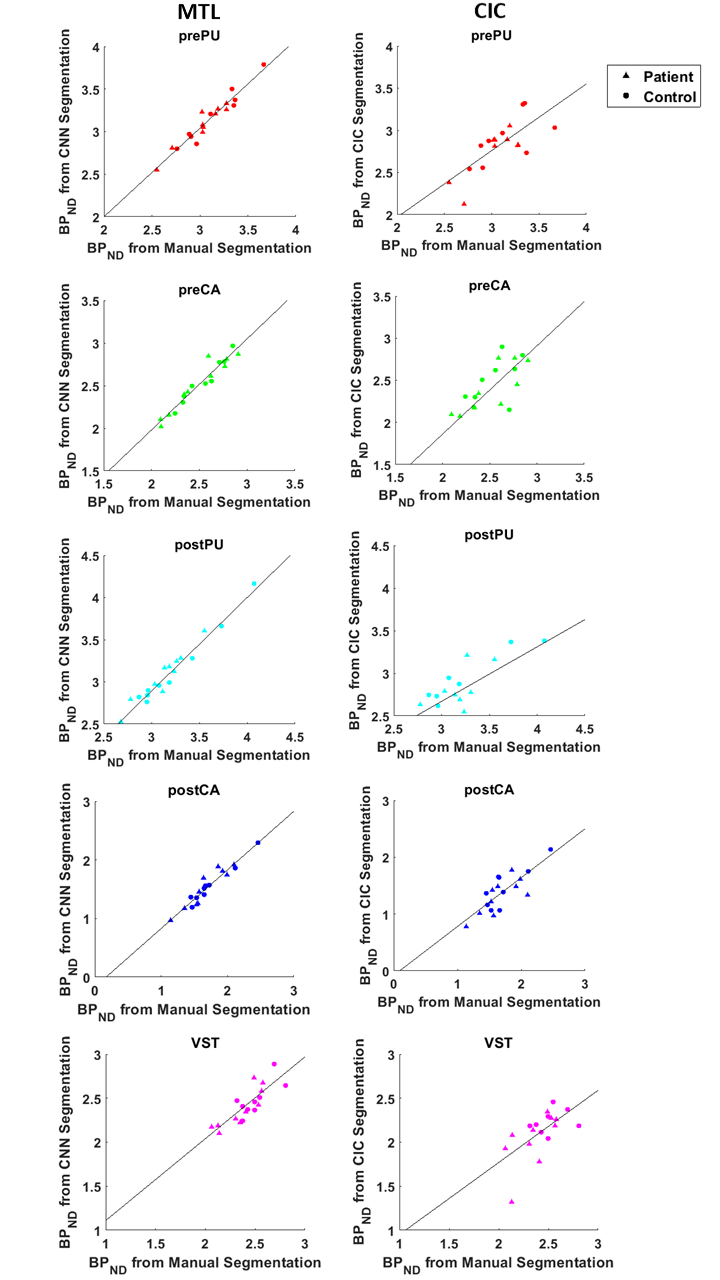
Figure 3. Scatter plots between BPND of the 5
subregions calculated using manual ROIs and both MTL- (left) and CIC-generated
(right) ROIs across 19 independent test subjects. Regression line for all
subregions is overlaid scatter plots, with regression coefficients found in
Table 2.