Bendik Skarre Abrahamsen1, Tone Frost Bathen1,2, Live Eikenes1, and Mattijs Elschot1,2
1Department of Circulation and Medical Imaging, NTNU, Trondheim, Norway, 2Department of Radiology and Nuclear Medicine, St. Olavs Hospital, Trondheim University Hospital, Trondheim, Norway
1Department of Circulation and Medical Imaging, NTNU, Trondheim, Norway, 2Department of Radiology and Nuclear Medicine, St. Olavs Hospital, Trondheim University Hospital, Trondheim, Norway
In PET/MR attenuation correction, direct estimation and correction for the bias between PET reconstructed with and without bone is a viable alternative to creating pseudo-CTs containing bone information.
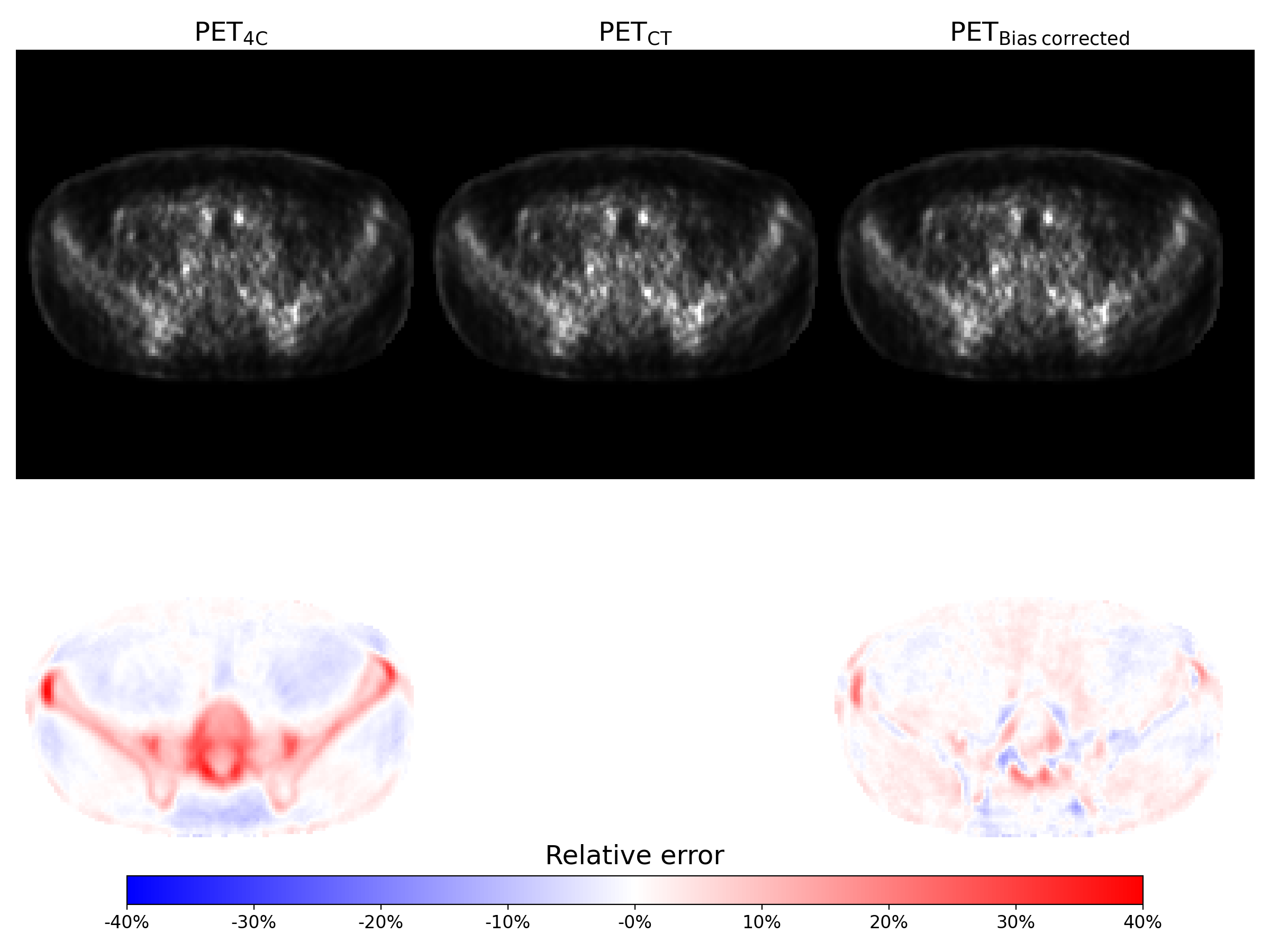
Figure 4 Top row: PET image reconstructed using the 4-class
umap, 4-class umap with bone information from CT and 4-class umap and corrected
using the generated bias image from left to right. All images are scaled the same.
Below are the relative error of each PET image compared to the PET
image reconstructed using the CT enhanced umap.
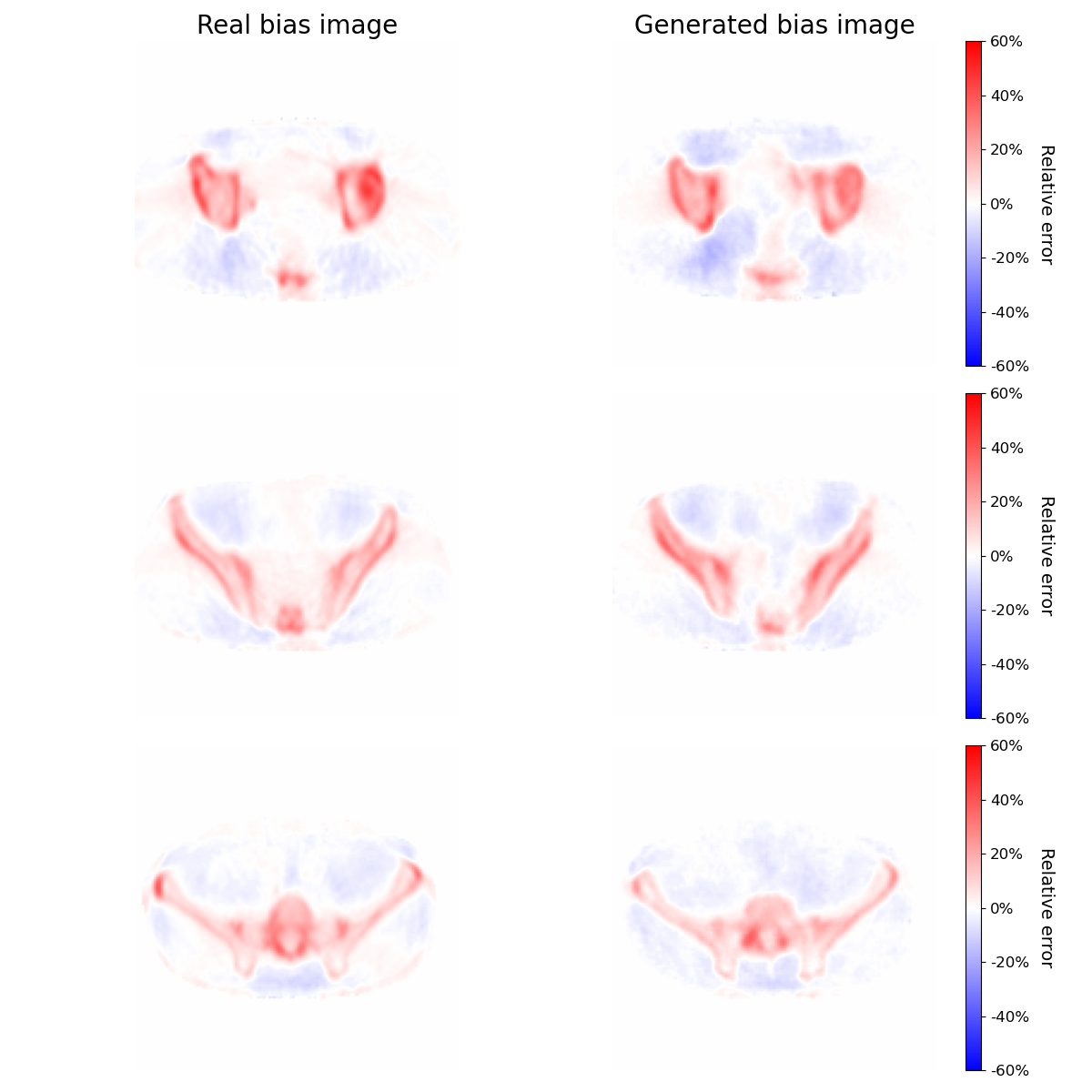
Figure 3: Real bias images and bias images generated by the pix2pix network. The bias image values correspond to the real and generated relative error between the PET image reconstructed using the 4-class attenuation correction map and the PET image reconstructed using the 4-class attenuation correction map with added bone information from a co-registered CT image. The images are from
the hold-out test set.