Radhika Madhavan1, Jaemin Shin2, Nastaren Abad1, Luca Marinelli1, J Kevin DeMarco3, Robert Y Shih3, Vincent B Ho3, Suchandrima Banerjee4, and Thomas K Foo1
1GE Global Research, Niskayuna, NY, United States, 2GE Healthcare, New York, NY, United States, 3Walter Reed National Military Medical Center and Uniformed Services University of the Health Sciences, Bethesda, MD, United States, 4GE Healthcare, Menlo Park, CA, United States
1GE Global Research, Niskayuna, NY, United States, 2GE Healthcare, New York, NY, United States, 3Walter Reed National Military Medical Center and Uniformed Services University of the Health Sciences, Bethesda, MD, United States, 4GE Healthcare, Menlo Park, CA, United States
Diffusion MRI often suffers from low signal-to-noise ratio, especially for high b-values. This work proposes a deep learning based denoising method to address this limitation, allowing the use of high b-values as well as higher spatial resolution.
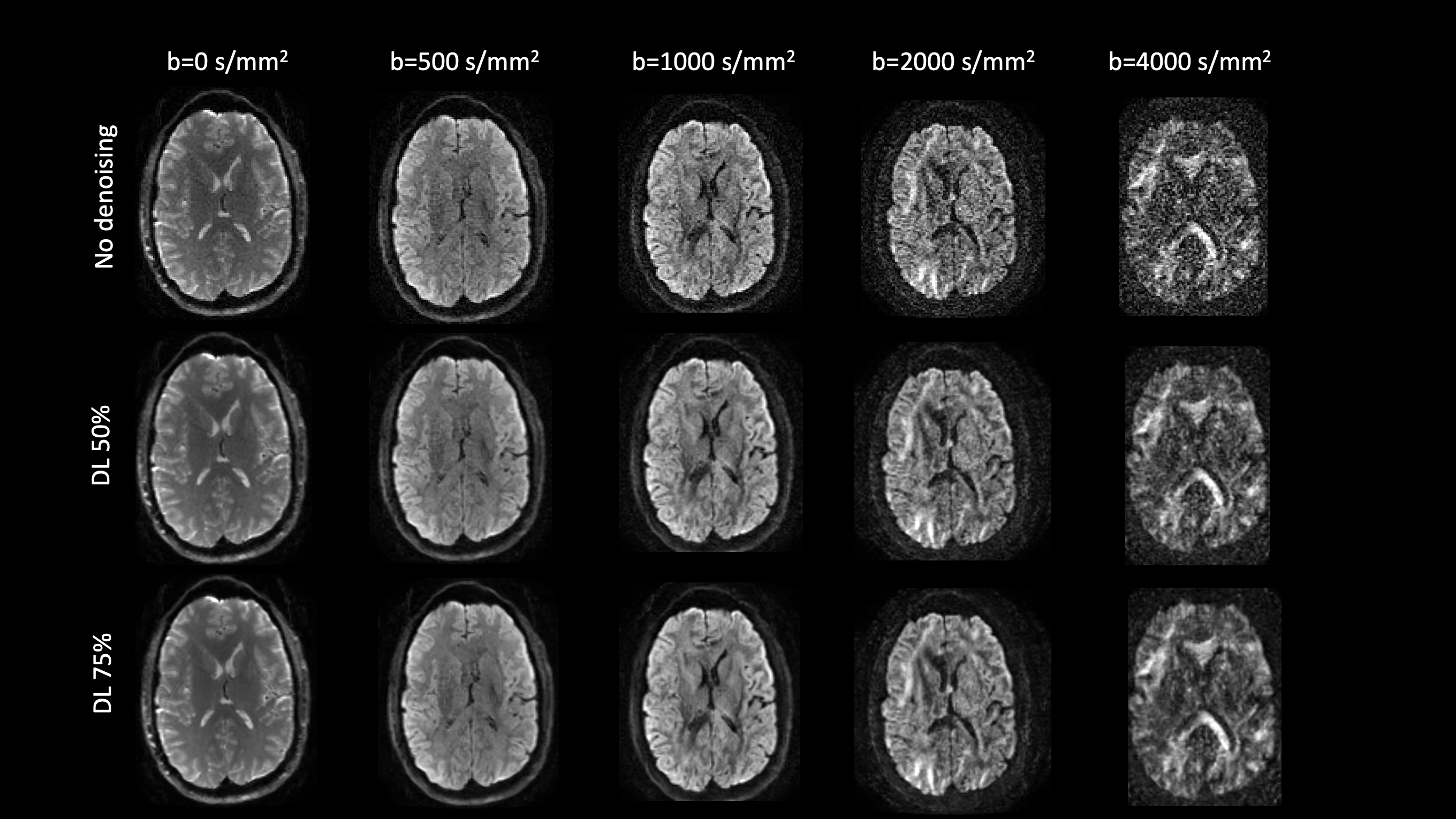
Figure 1: Diffusion weighted images for an example healthy volunteer showing improved SNR (separability of gray and white matter compartments in b-values >=2000 s/mm2) without spatial smoothing across multiple intensities of DL-based denoising.
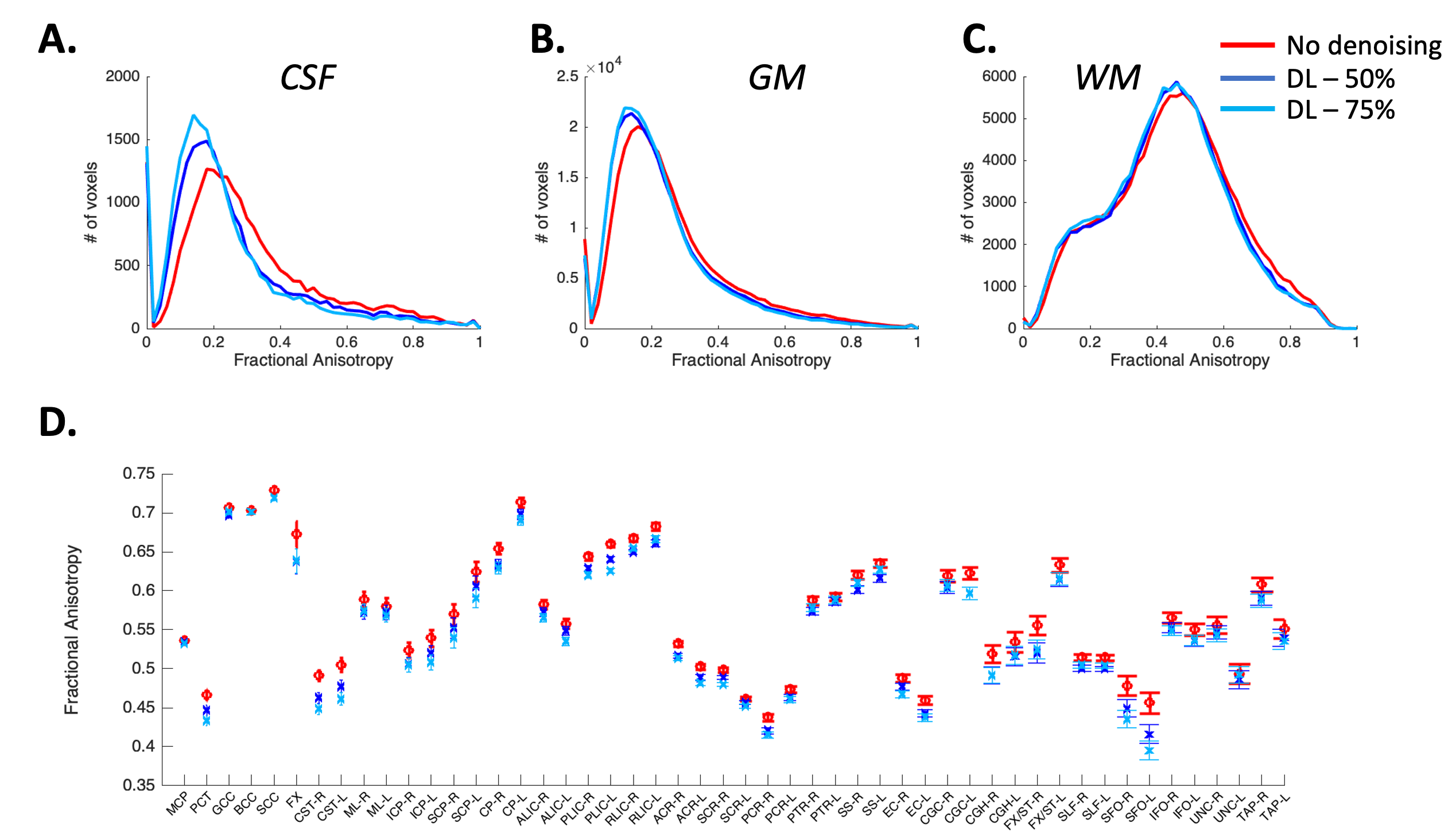
Figure 3: DL-based denoising improved differentiation across signal compartments while preserving quantitative tensor-based metrics. (A-C) Histogram analysis demonstrating distribution of FA values in whole brain CSF, Gray matter (GM) and WM. Quantitative diffusion metrics were compared across two level of DL denoising (50% and 75%). Results were consistent across both volunteers and across acquisitions. (D) FA values across 50 WM regions remain unchanged demonstrating that DL-based denoising maintains tensor-based quantitative metrics.