Yuhao Yan1,2 and Zheng Chang1,2
1Medical Physics Graduate Program, Duke University, Durham, NC, United States, 2Department of Radiation Oncology, Duke University, Durham, NC, United States
1Medical Physics Graduate Program, Duke University, Durham, NC, United States, 2Department of Radiation Oncology, Duke University, Durham, NC, United States
Neural networks can
accelerate brain DTI. Cascade-net out-performed U-net and PD-net, obtaining comparable
image quality as compared with the reference reconstructed from the full
k-space data on reconstruction of DTI images, ADC maps and FA maps.
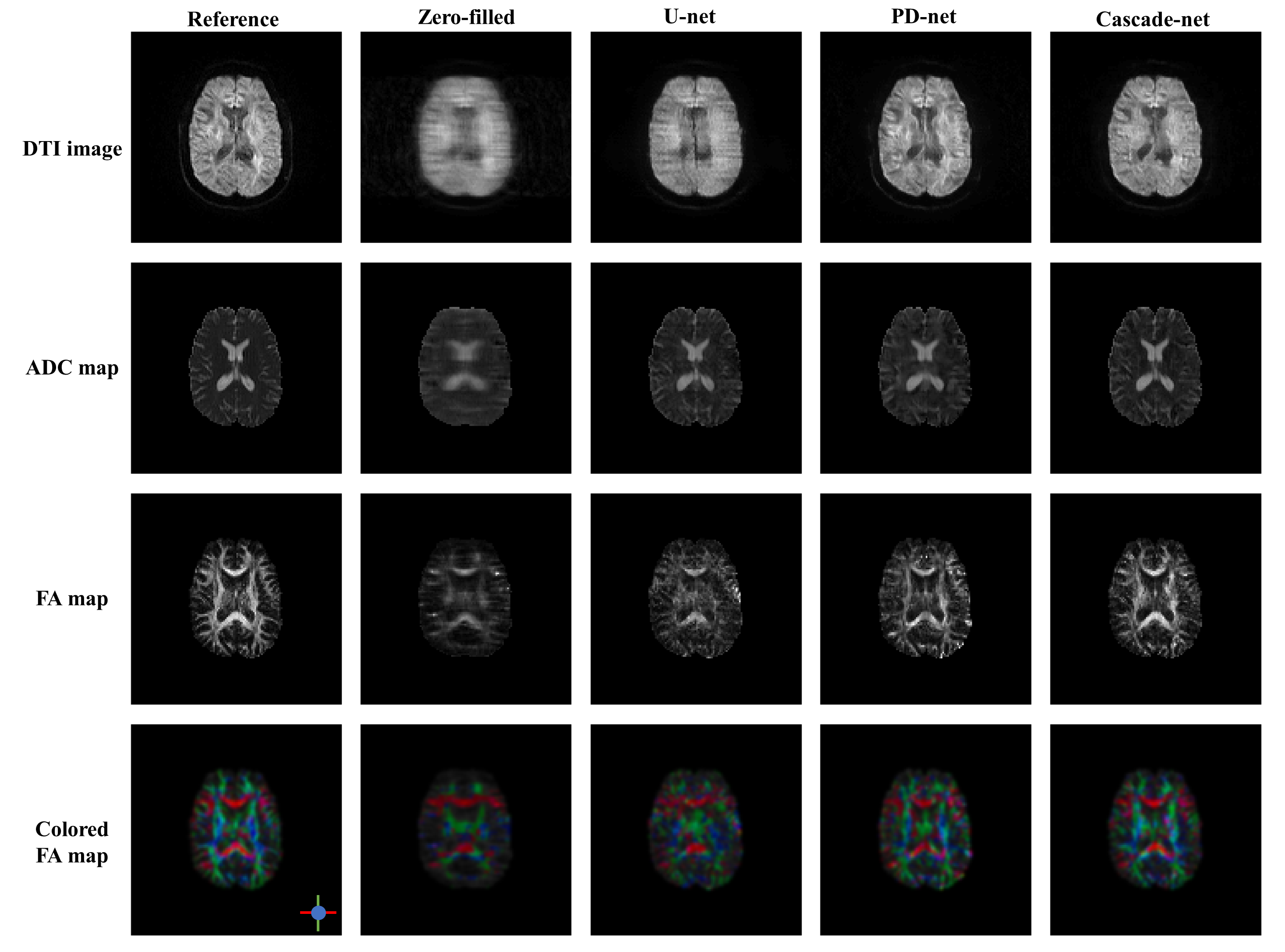
Figure 3. Illustration of DTI images, ADC maps, FA maps and colored FA maps of a
selected slice, from top to bottom row. Each column from left to right are
reference images reconstructed from full k-space data, images reconstructed
from zero-filled under-sampled k-space data, images reconstructed from
under-sampled k-space data using U-net, PD-net and Cascade-net.
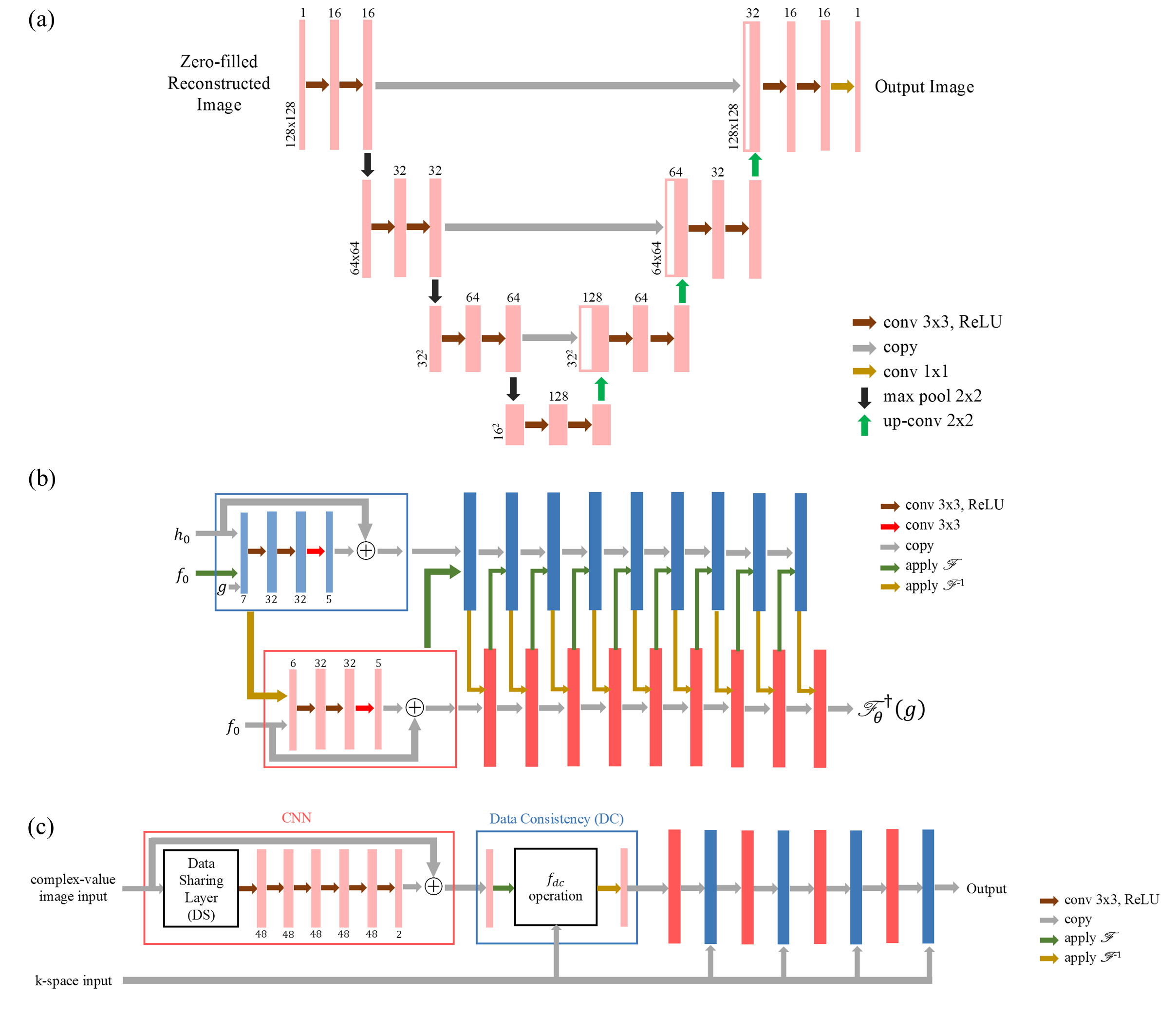
Figure 1.a. Structure of U-net.1,4 The number of filters in each layer is a quarter of the original
structure. Zero-padding is adapted to maintain the size of images in
convolutions. 1.b. Structure of PD-net.5 In this work,
the activation function PReLU non-linearity in the original structure was
substituted by ReLU non-linearity for consistency with U-net and Cascade-net.1
1.c. Structure of Cascade-net.2 The number of
filters was set as 48 in this work, which was 64 in the original structure.1