Emmanuelle Weber1, Christoph Leuze1, Daniel A. N. Barbosa1, Gustavo Chau Loo Kung1, Kalanit Grill-Spector1, and Jennifer A. McNab1
1Stanford, Stanford, CA, United States
1Stanford, Stanford, CA, United States
Feasibility
study of a machine learning direct prediction of tissue microstructure from raw
diffusion MRI data. We attempted to predict the well-understood main fiber
orientation from both simulated and dMRI-3D histology dataset.
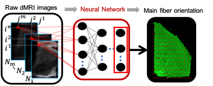
Figure 1: Deep learning
framework aiming at predicting the microstructural features from raw diffusion
MRI (dMRI) data using histology images as ground truth. Example of prediction
of main fiber orientation from previously acquired data on human thalamus.
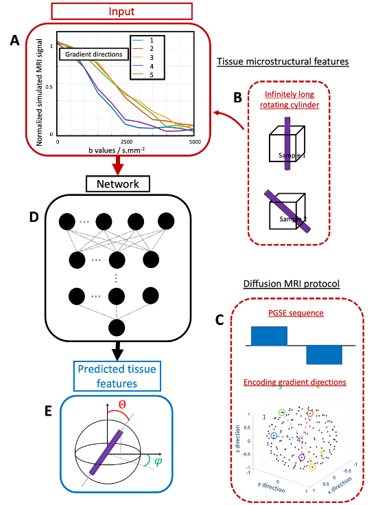
Figure
2: Summary of the whole pipeline that enables to
predict a single simulated fiber orientation using deep learning. A) Normalized
dMRI signal from an B) infinitely long rotating cylinder as a function of the
C) PGSE sequence b values for different gradient orientation. D) This signal is
then fed to a neural network to predict the E) orientation of the cylinder
given by the spherical angles theta and phi.