Julian Rauch1,2, Dominik Ludwig1,2, Frederik B. Laun3, and Tristan A. Kuder1
1Division of Medical Physics in Radiology, German Cancer Research Center (DKFZ), Heidelberg, Germany, 2Faculty of Physics and Astronomy, Heidelberg University, Heidelberg, Germany, 3Institute of Radiology, University Hospital Erlangen, Friedrich-Alexander-Universität Erlangen-Nürnberg (FAU), Erlangen, Germany
1Division of Medical Physics in Radiology, German Cancer Research Center (DKFZ), Heidelberg, Germany, 2Faculty of Physics and Astronomy, Heidelberg University, Heidelberg, Germany, 3Institute of Radiology, University Hospital Erlangen, Friedrich-Alexander-Universität Erlangen-Nürnberg (FAU), Erlangen, Germany
The
signal stability of AXR measurements is slightly improved when suppressing
pulsation-induced variations by ECG triggering. However, pulsation does not
seem to be the main source of signal variations.
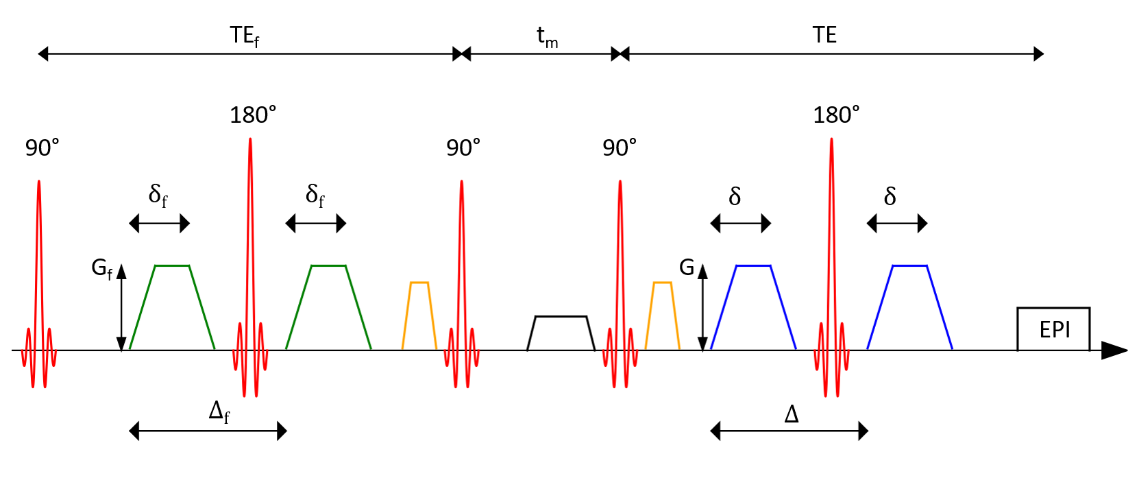
Figure
1: Schematic
representation of a filter exchange imaging (FEXI) sequence using two pulsed
gradient spin echo (PGSE) blocks. The first gradient pair used as the FEXI
filter is followed by a varying mixing time during which
the magnetization is longitudinally stored while transversal components are
dephased. Before and after the second and third radiofrequency pulse,
respectively, gradients to choose the right echo path are applied. The second
gradient pair is a standard diffusion weighting. This block is followed by an
echo planar imaging (EPI) readout.
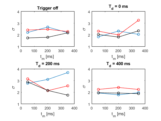
Figure
2: Comparison of the standard deviations σ resulting from the different trigger
experiments. No diffusion weighting was applied (b = 0 s/mm2).
The three used
orthogonal diffusion encoding directions (2/3, 2/3, 1/3), (1/3, 2/3, 2/3) and (2/3, 1/3, 2/3)
are depicted in blue,
red and black, respectively.