Ratthaporn Boonsuth1, Marco Battiston1, Francesco Grussu1,2, Marios Yiannakas1, Torben Schneider3, Rebecca Samson1, Ferran Prados1,4,5, and Claudia A. M. Gandini Wheeler-Kingshott1,6,7
1NMR research Unit, Queen Square MS Centre, Department of Neuroinflammation, UCL Queen Square Institute of Neurology, London, United Kingdom, 2Radiomics Group, Vall d’Hebron Institute of Oncology, Vall d’Hebron Barcelona Hospital Campus, Barcelona, Spain, 3Philips Healthcare, Guildford, Surrey, United Kingdom, 4Centre for Medical Image Computing, Medical Physics and Biomedical Engineering, University College London, London, United Kingdom, 5E-Heath Centre, Universitat Oberta de Catalunya, Barcelona, Spain, 6Department of Brain & Behavioural Sciences, University of Pavia, Pavia, Italy, 7Department of Brain Connectivity Centre Research Department, IRCCS Mondino Foundation, Pavia, Italy
1NMR research Unit, Queen Square MS Centre, Department of Neuroinflammation, UCL Queen Square Institute of Neurology, London, United Kingdom, 2Radiomics Group, Vall d’Hebron Institute of Oncology, Vall d’Hebron Barcelona Hospital Campus, Barcelona, Spain, 3Philips Healthcare, Guildford, Surrey, United Kingdom, 4Centre for Medical Image Computing, Medical Physics and Biomedical Engineering, University College London, London, United Kingdom, 5E-Heath Centre, Universitat Oberta de Catalunya, Barcelona, Spain, 6Department of Brain & Behavioural Sciences, University of Pavia, Pavia, Italy, 7Department of Brain Connectivity Centre Research Department, IRCCS Mondino Foundation, Pavia, Italy
The
diffusion-derived metrics are reproducible, with no significant differences
between pre- and post-upgrade in spinal cord, white matter, and grey matter.
Following a major scanner upgrade at a single site, diffusion measurements were
found not to differ substantially
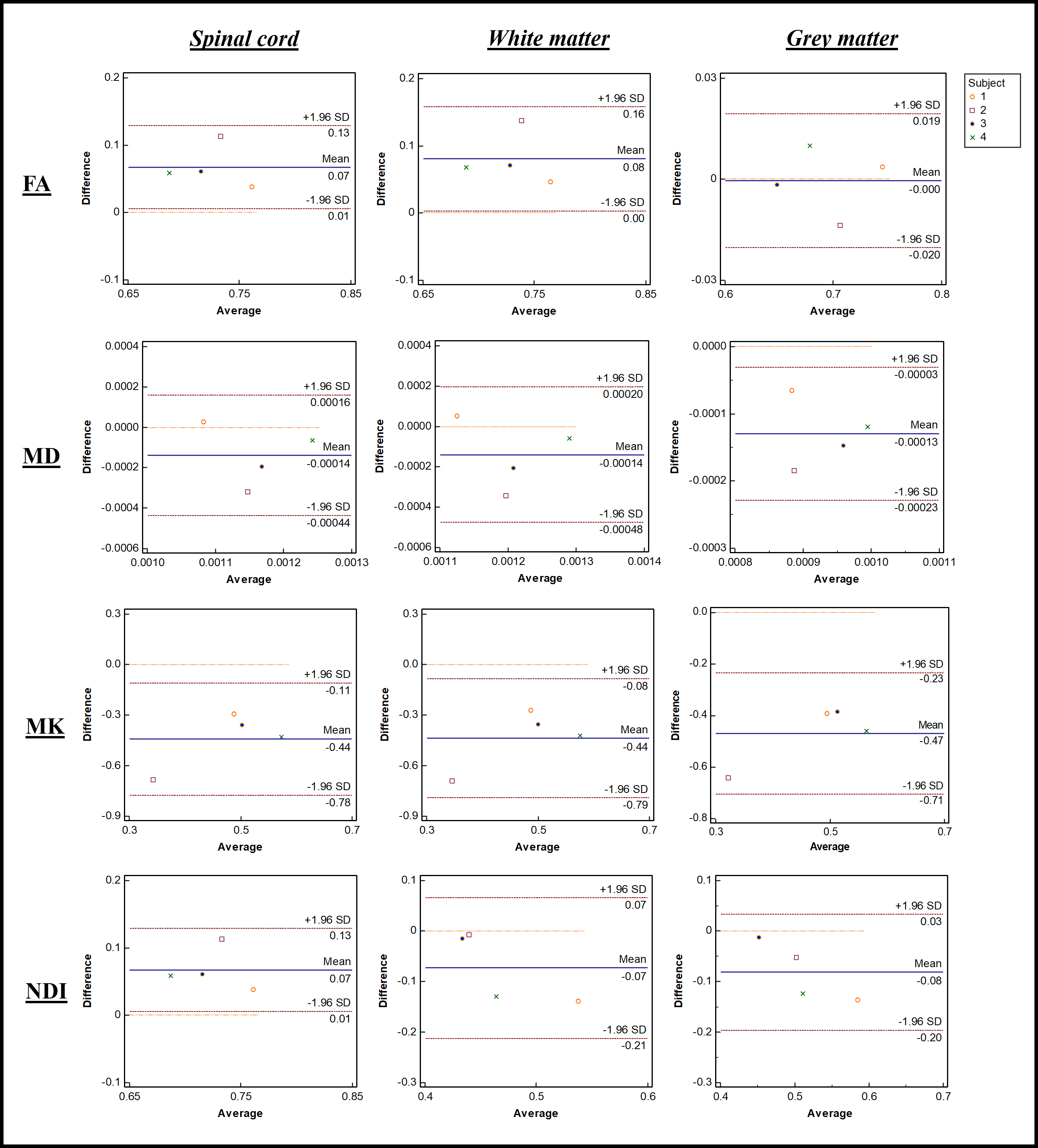
Figure 1.
Bland–Altman plots of four diffusion metrics for 3 different ROIs, namely whole
spinal cord area, white matter, and grey matter. The x and y-axes represent,
respectively, average, and mean differences between pre- and post-upgrade. The
blue lines show the mean of the differences, while the pairs of dotted red
lines denote the 95% confidence intervals.
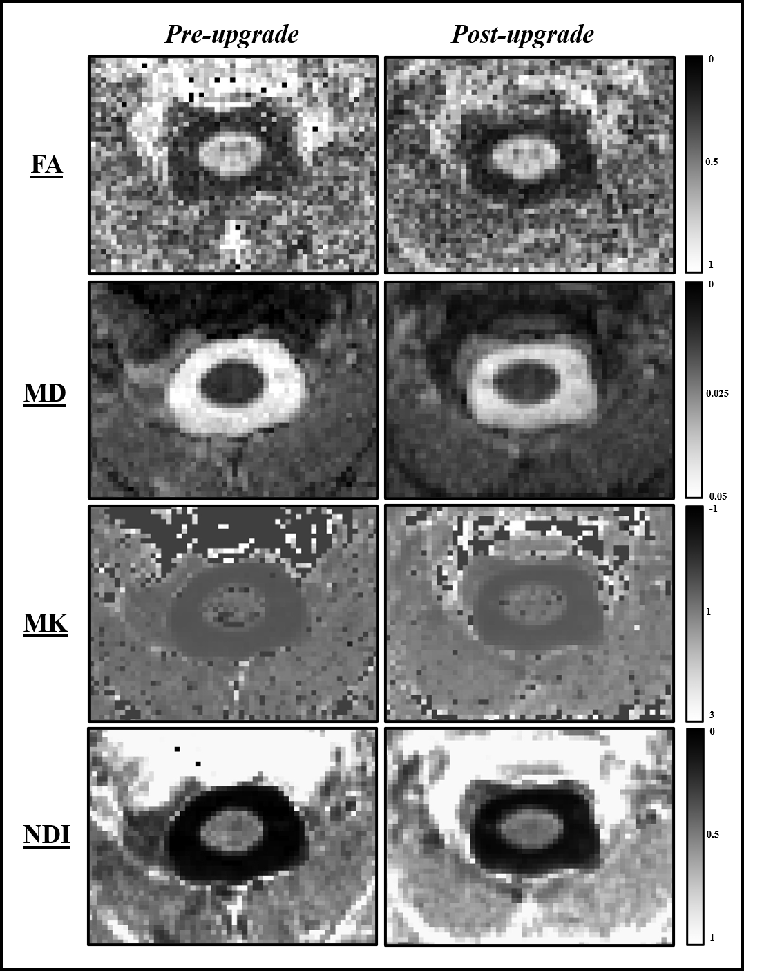
Figure 2. Parametric maps for fractional anisotropy (FA),
mean diffusity (MD), mean kurtosis (MK) and neurite density index (NDI) from
a single subject obtained pre- and post-upgrade.