Samo Lasic1,2 and Henrik Lundell1
1Danish Research Centre for Magnetic Resonance, Centre for Functional and Diagnostic Imaging and Research, Copenhagen University Hospital Hvidovre, Copenhagen, Denmark, 2Random Walk Imaging, Lund, Sweden
1Danish Research Centre for Magnetic Resonance, Centre for Functional and Diagnostic Imaging and Research, Copenhagen University Hospital Hvidovre, Copenhagen, Denmark, 2Random Walk Imaging, Lund, Sweden
Ellipsoidal tensor encoding with independent control of spectral anisotropy and tuning yields distinctly different signal signatures for compartments with different anisotropic time-dependent diffusion.
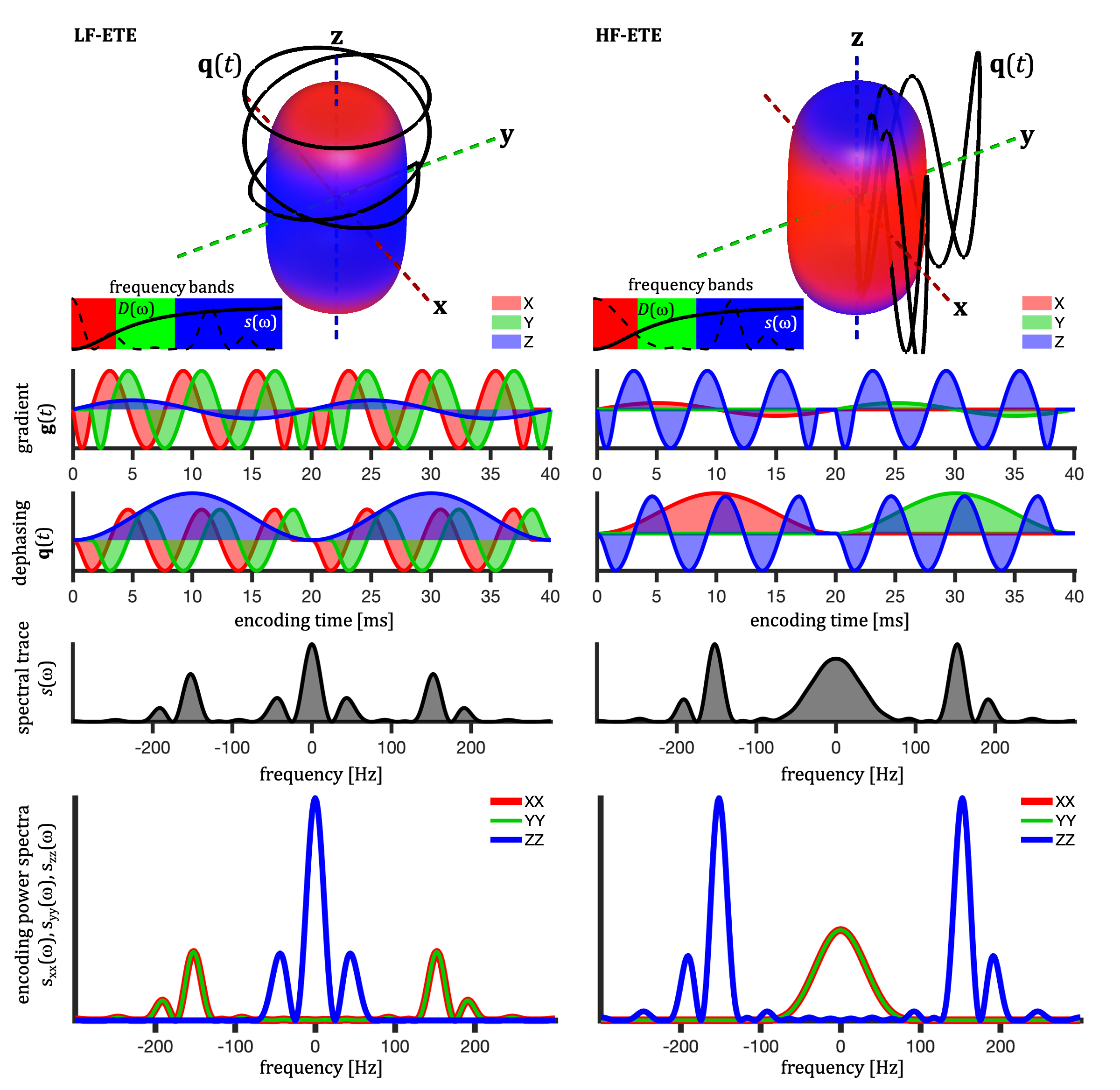
Figure 1: Tuned ellipsoidal tensor encodings with varying spectral anisotropy featuring more encoding power at low frequencies (LF-ETE) or high frequencies (HF-ETE) along z-axis. While the trace spectra is similar for LF-ETE and HF-ETE, the power distribution across different directions is markedly different. Color coding is based on the RGB-weights given by projections of the encoding power in the three bands with crossover frequencies determined by $$$D(\omega)$$$ for a sphere with $$$D_0^2R^{-4}=8000 \,\mathrm{s}^{-2}$$$ reaching 1/3 and 2/3 of the asymptotic value.
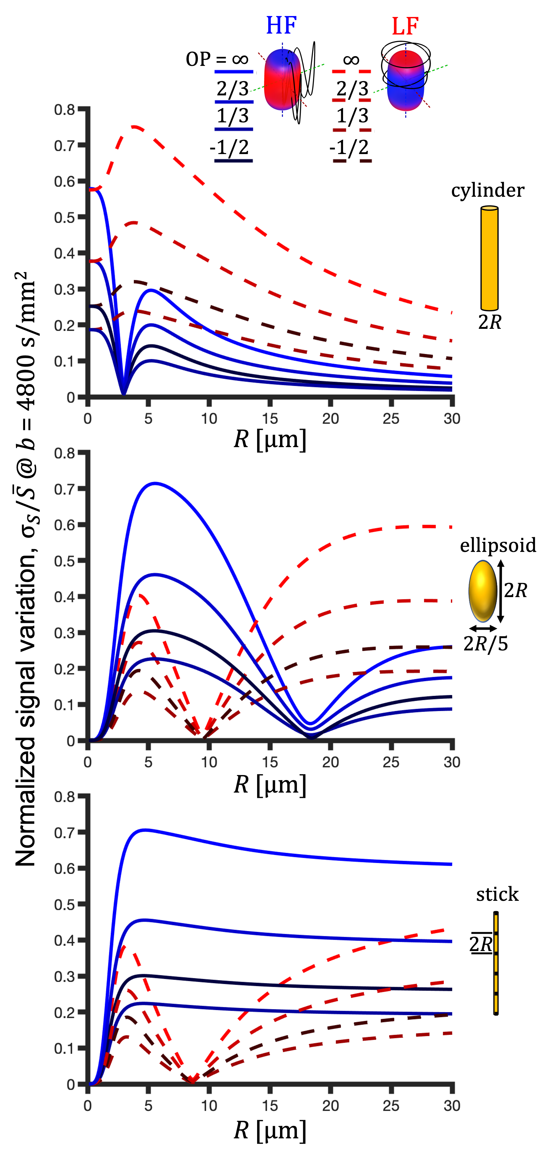
Figure 3: Directional spread of signals (normalized STD, $$$\sigma_\mathrm{S}/\bar{S}$$$) as a function of compartment size for cylindrical, ellipsoidal and stick-like restrictions. HF-ETE (solid blue lines) and LF-ETE (dashed red lines) for Watson orientation distributions with varying order parameters (OP). Location of the minima is independent of OP. For cylindrical restrictions, the minima occur only with the HF-ETE, while for stick-like restrictions, the minima occur only with the LF-ETE. For ellipsoidal restrictions we have minima with both LF-ETE and HF-ETE.