Kristin Koller1, Chantal MW Tax1,2, Dmitri Sastin1,3,4, and Derek K Jones1,5
1Cardiff University Brain Research Imaging Centre, Cardiff, United Kingdom, 2Image Sciences Institute, University Medical Center Utrecht, Utrecht, Netherlands, 3Department of Neurosurgery, University Hospital of Wales, Cardiff, United Kingdom, 4BRAIN Biomedical Research Unit, Health & Care Research Wales, Cardiff, United Kingdom, 5Mary MacKillop Institute for Health Research, Australian Catholic University, Melbourne, Australia
1Cardiff University Brain Research Imaging Centre, Cardiff, United Kingdom, 2Image Sciences Institute, University Medical Center Utrecht, Utrecht, Netherlands, 3Department of Neurosurgery, University Hospital of Wales, Cardiff, United Kingdom, 4BRAIN Biomedical Research Unit, Health & Care Research Wales, Cardiff, United Kingdom, 5Mary MacKillop Institute for Health Research, Australian Catholic University, Melbourne, Australia
High test-retest repeatability of microscopic FA (μFA) and isotropic and anisotropic diffusional variance (MKi and MKa) derived from free gradient waveforms is demonstrated in a cohort of 6 participants scanned 5 times on an ultra-high gradient MRI scanner.
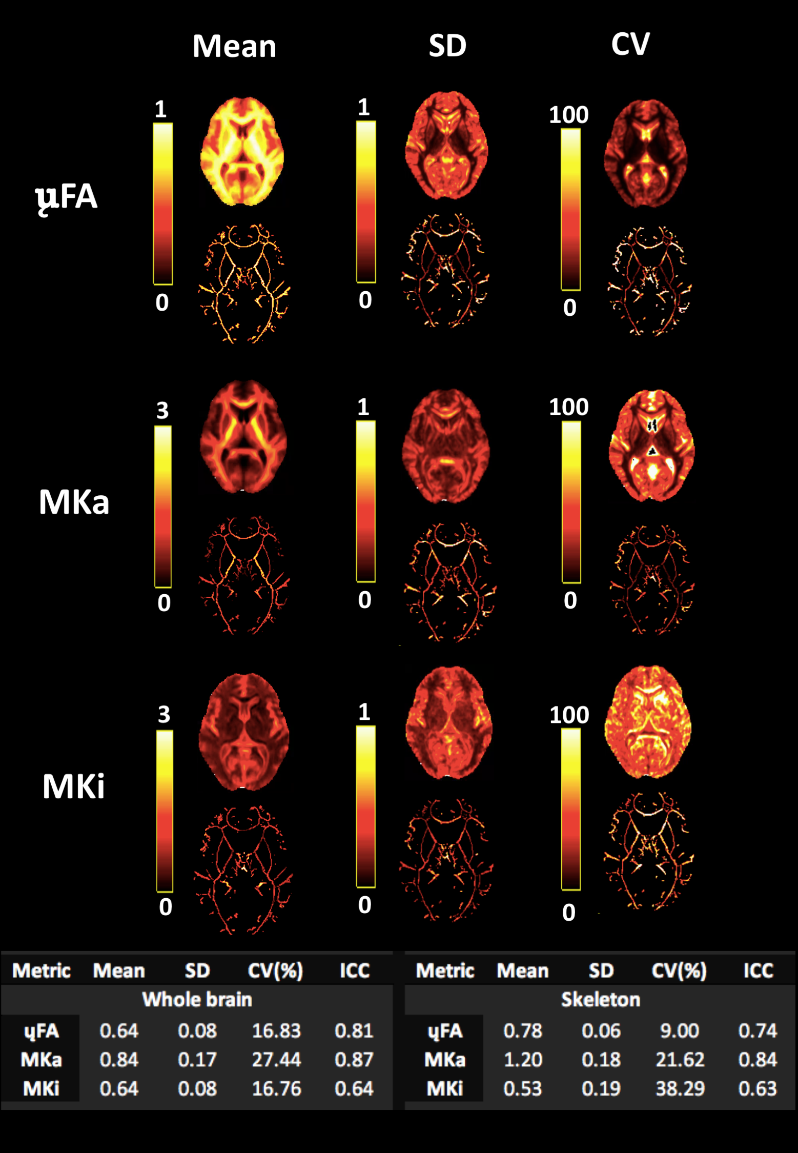
Fig. 3. Summary statistical maps for each participant averaged across all sessions. μFA. As expected, the bright voxels are highest in dense white matter regions. Bright voxels in SD and COV maps show more variability in regions contaminated with cerebrospinal fluid such as the ventricles, and low variability in white matter regions. MKi. Bright voxels located in areas of isotropic diffusion such as CSF in the ventricles. MKa Bright voxels demonstrate regions of strong anisotropic diffusion.
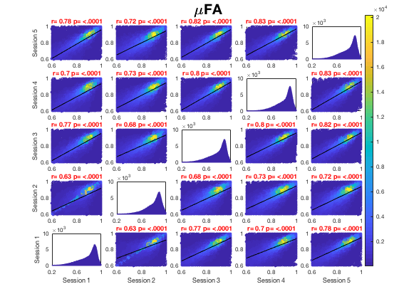
Fig. 2A. Voxel-wise whole brain white matter Pearson correlations are presented between individual time points for μFA white matter skeletons. Voxels pooled across all subjects for each map, r = Pearson correlation coefficient, p<.0001 for all plots. Univariate histograms on the diagonal show distributions of voxels of μFA across all voxels. Calculation of metrics in session 2 of one subject was not robust and thus excluded.