Kentaro Akazawa1, Koji Sakai1, Tomoaki Kitaguchi1, Tomonori Toyotsuji1, Thorsten Feiweier 2, Hiroshi Imai3, and Kei Yamada1
1Radiology, Kyoto Prefectural University of Medicine, Kyoto, Japan, 2Siemens Healthcare GmbH, Erlangen, Germany, 3Siemens Healthcare K.K., Shinagawa, Japan
1Radiology, Kyoto Prefectural University of Medicine, Kyoto, Japan, 2Siemens Healthcare GmbH, Erlangen, Germany, 3Siemens Healthcare K.K., Shinagawa, Japan
A relatively
short mixing time of 30 msec for double diffusion encoding is likely adequate
to evaluate the microscopic fractional anisotropy not only in the normal brain
structures, but also in pathologically abnormal areas such as brain tumors.
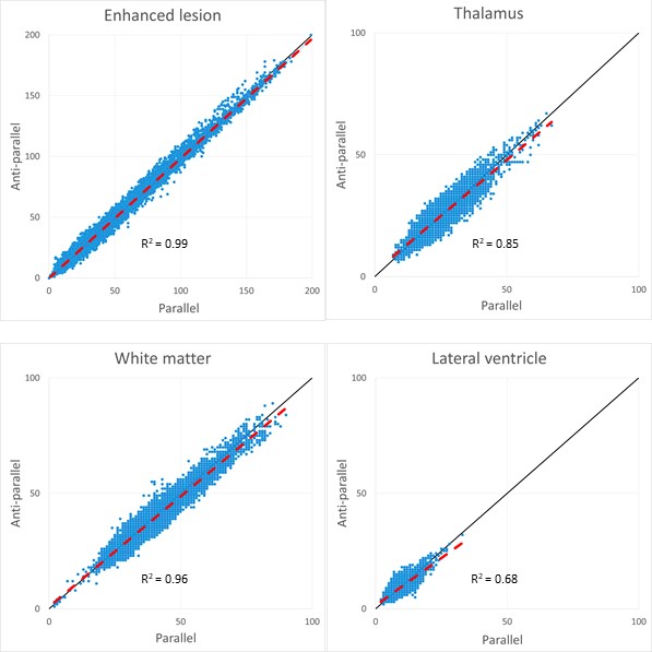
The scatter
plots of relative signal intensities in the enhanced lesions in brain tumors,
the normal thalamus, the normal white matters, and the lateral ventricle. The
horizontal and vertical axes are relative signal intensities from the parallel
directions and from the anti-parallel directions, respectively. There were
significantly correlated in all areas (p
< 0.001).
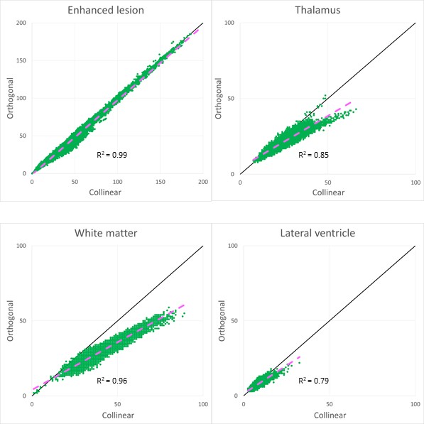
The
scatter plots of relative signal intensities in the enhanced lesions, the normal
thalamus, the normal white matters, and the lateral ventricle. The horizontal
and vertical axes are relative signal intensities from the collinear directions
which are the averages of the parallel and anti-parallel directions, and from
the orthogonal directions, respectively. There were significantly correlated in
all areas (p < 0.001). The slopes
of the thalamus and white matter seemed to be different from those in figure 2.