Rafael N Henriques1, Sune N. Jespersen2,3, Derek K. Jones4,5, and Jelle Veraart6
1Champalimaud Research, Champalimaud Centre for the Unknown, Lisbon, Portugal, 2Department of Clinical Medicine, Aarhus University, Aarhus, Denmark, 3Department of Physics and Astronomy, Aarhus University, Aarhus, Denmark, 4School of Psychology, Cardiff University, Cardiff, United Kingdom, 5Mary MacKillop Institute for Health Research, Australian Catholic University, Melbourne, Australia, 6Center for Biomedical Imaging, NYU Grossman School of Medicine, New York, NY, United States
1Champalimaud Research, Champalimaud Centre for the Unknown, Lisbon, Portugal, 2Department of Clinical Medicine, Aarhus University, Aarhus, Denmark, 3Department of Physics and Astronomy, Aarhus University, Aarhus, Denmark, 4School of Psychology, Cardiff University, Cardiff, United Kingdom, 5Mary MacKillop Institute for Health Research, Australian Catholic University, Melbourne, Australia, 6Center for Biomedical Imaging, NYU Grossman School of Medicine, New York, NY, United States
Our novel regularized DKI estimator improves the robustness and reproducibility of the kurtosis metrics and results in parameter maps with enhanced quality and contrast; thereby promoting the wider use of DKI in clinical research and potentially diagnostics
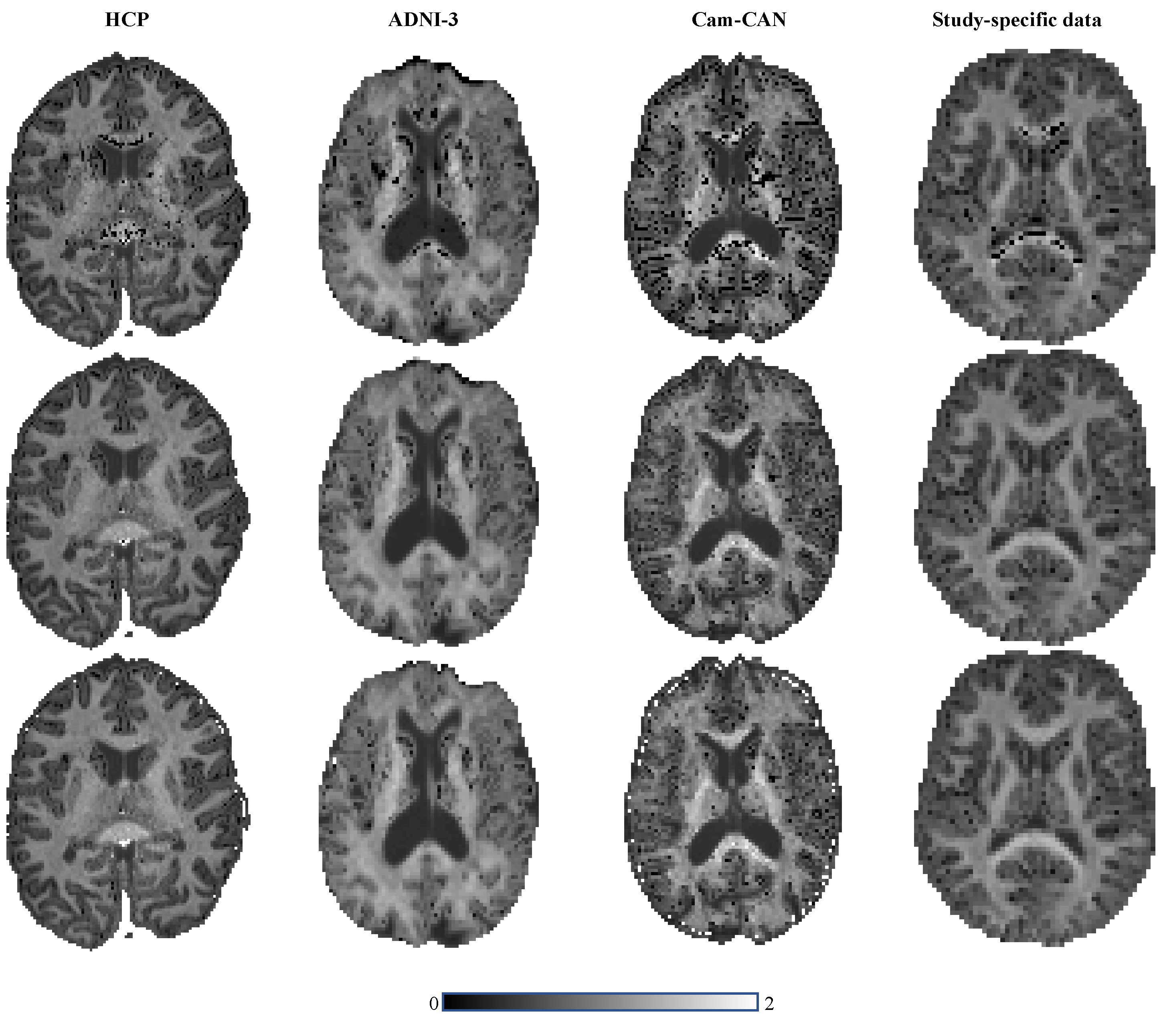
Figure 4: The $$$\bar{K}$$$ maps for the various data set are shown for the ordinary and regularized NLS in the top and middle row, respectively. Moreover, we show the map of the predicted mean kurtosis $$$\hat{K}$$$ (bottom row) to illustrate the similarity in contrast.
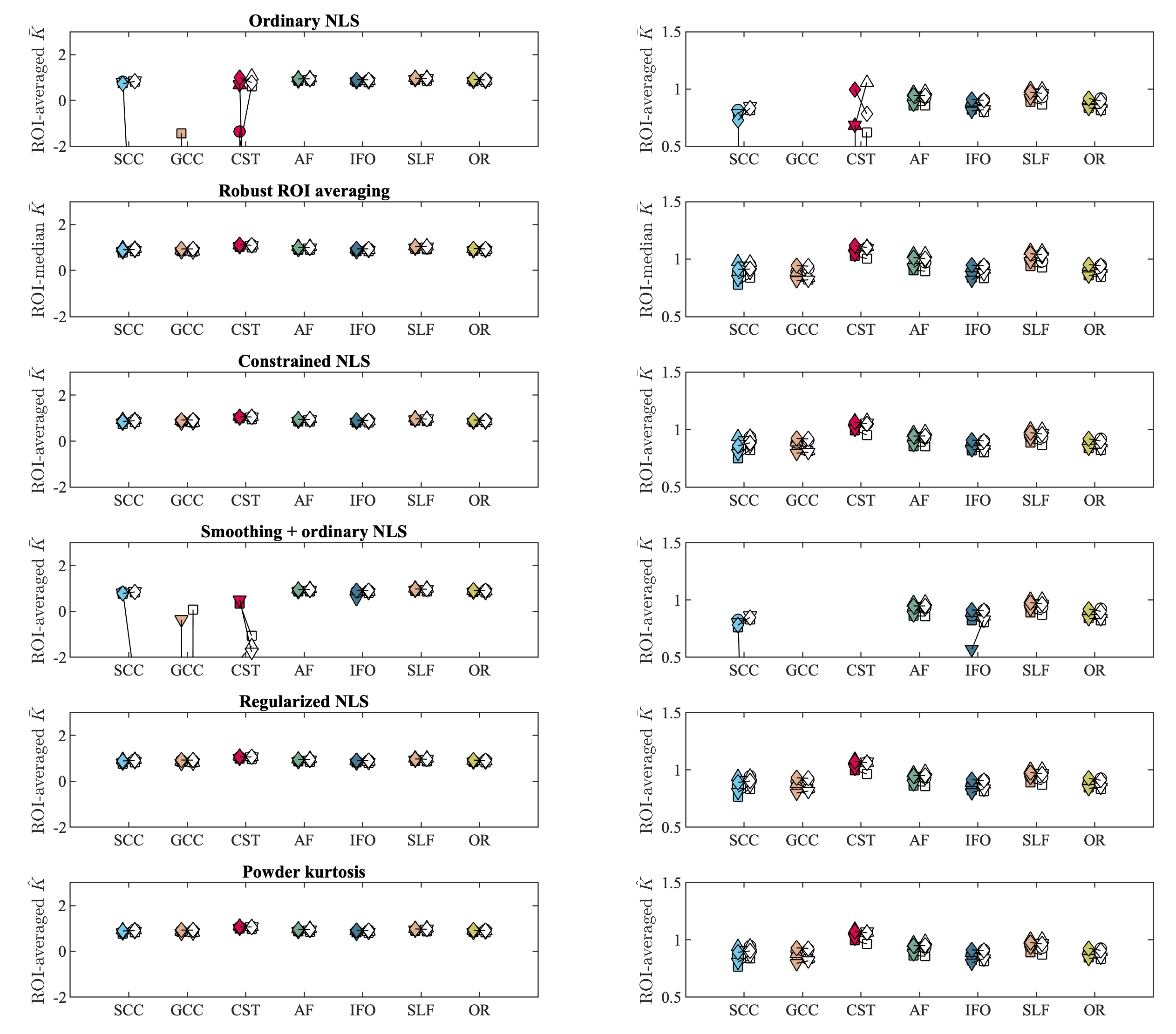
Figure 2: The tract-averaged $$$\bar{K}$$$ using various fitting strategies and $$$\hat{K}$$$ (bottom row) for the test (filled marker) and retest data (open marker) each subject (labeled by marker shape). The graphs on the right column show the same data, but windowed differently for enhanced contrast. Seven major white matter tracts were evaluated: genu and splenium of the corpus callosum (GCC and SCC), corticospinal tract (CST), arcuate fasciculus (AF), inferior fronto-occipital fasciculus (IFO), Superior Longitudinal Fasciculus (SLF) and optic radiation (OR).